Neural Circuitry and Neurotransmitters
That Mediate the Acoustic Startle Reflex
That Mediate the Acoustic Startle Reflex
Michael Davis, mdavis4@emory.edu
Emory University School of Medicine
Atlanta, Georgia, USA
Popular version of paper 3pAB2
Presented Wednesday afternoon, November 3, 1999
138th ASA Meeting, Columbus, Ohio
When we hear a loud, abrupt sound, we often startle. In fact, all mammals tested so far display various components of a startle reaction when presented with such stimuli. The response consists of a rapid extension and then flexion of a series of muscles. Although the startle response is a reflex that is usually not under voluntary control, it can be modified by many different treatments. For example, people or rats have bigger startle reactions when they are afraid. This increase can be reduced by medications that reduce anxiety. Near threshold stimuli presented shortly before a loud startle producing sound reduce the size of the startle reflex. This effect is abnormal in certain types of psychiatric patients. Hence, changes in the startle reflex are now widely used to study how the brain generates fear, anxiety, or gates irrelevant sensory information.

In humans, changes in electrical activity in neck muscles can occur within 9 msec after the onset of an auditory stimulus and within 14 msec in jaw muscles. In rats, startle occurs within 5 msec in the neck and 8 msec in the hindleg. Because the acoustic startle reflex has such a short latency, a simple neural pathway must mediate it. In 1982 our laboratory proposed that acoustic startle in the rat was mediated by four synapses: three in the brainstem (the ventral cochlear nucleus, an area to the middle and just below the ventral nucleus of the lateral lemniscus, and the nucleus reticularis pontis caudalis) and one synapse onto motoneurons in the spinal cord. Electrolytic lesions of these nuclei eliminated acoustic startle while single pulse electrical stimulation of these nuclei elicited startle-like responses with a progressively shorter latency (less lag time between stimulus and response) as the electrode was moved farther down the startle pathway. However, we now have found that selective destruction of cell bodies without a concomitant loss of fibers of passage in the ventral nucleus of the lateral lemniscus or the area to the middle and just below it did not eliminate startle whereas comparable lesions in the nucleus reticularis pontis caudalis still completely eliminated startle. These data indicate that an additional synapse in the ventral nucleus of the lateral lemniscus or the area to the middle and just below it may not be necessary for relaying auditory information from the cochlear nucleus to the pontine reticular formation. On the other hand, all the data still pointed to the critical importance of the nucleus reticularis pontis caudalis in the acoustic startle reflex.
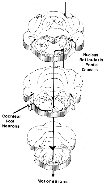
Until very recently, it has been unclear how auditory information gets to this traditionally non-auditory part of the brainstem. In both rodents and humans there are a small group of very large cells (35 m in diameter) embedded in the cochlear nerve, called cochlear root neurons. These neurons receive direct input from the spiral ganglion cells in the cochlea, making them the first acoustic neurons in the central nervous system. They send exceedingly thick axons (sometimes as wide as 7 m) through the trapezoid body, at the very base of the brain, directly to an area to the middle and just below the lateral lemniscus and continue on up to the deep layers of the superior colliculus. However, they give off thick axon collaterals that terminate directly in the nucleus reticularis pontis caudalis, exactly at the level known to be critical for the acoustic startle reflex. Most importantly, chemical destruction of these cochlear root neurons completely eliminates the acoustic startle reflex. Although damage to the auditory root, where the cochlear root neurons reside, has not been fully ruled out, other tests indicated that these animals could clearly orient to auditory stimuli (e.g. suppression of licking) and had normal compound action potentials recorded from the cochlear nucleus.
Hence, we believe the acoustic startle pathway may consist of only three synapses onto 1) cochlear root neurons; 2) neurons in the nucleus reticularis pontis caudalis and 3) motoneurons in the facial motor nucleus (pinna reflex and perhaps the eyeblink) or spinal cord (whole body startle - see figure above).
In humans, the obicularis oculi muscles innervated by the facial motor nucleus mediate the eyeblink component of startle. Because the facial motor nucleus receives a direct projection from the nucleus reticularis pontis caudalis, the eyeblink component of startle could involve a pathway consisting of cochlear root neurons, the nucleus reticularis pontis caudalis and the facial motor nucleus. However, this simple neural pathway may not be able to account for the relatively long latency of the eyeblink reflex (35 msec) so that other pathways have been suggested.