[ Lay Language Paper Index | Press Room ]
Peter J. Kaczkowski - peter@apl.washington.edu
Shahram Vaezy, Roy Martin, and Lawrence Crum
Center for Industrial and Medical Ultrasound
Applied Physics Laboratory
University of Washington
Seattle, WA 98105
George Keilman
Sonic Concepts
Woodinville, WA 98072-7029
Ultrasound energy has been used for decades to safely image the interior of the body without the risk associated with ionizing radiation such as X-rays, and at much lower cost than Magnetic Resonance Imaging (MRI). Medical imaging with ultrasound is done at very low power and the risk of any damage to tissue is extremely small. At higher powers, however, ultrasound can be used in therapeutic and surgical applications, principally by depositing heat at highly targeted locations within the body. Medical ultrasound is a high frequency mechanical wave that moves matter slightly as it passes through it, compressing and expanding tissue millions of times a second. Heat is generated by the process because all materials absorb a tiny fraction of the energy carried by the ultrasonic wave through friction of motion, and at very high powers this absorption can lead to a dramatic rise in temperature.
Ultrasound can be highly focused, as evidenced by the sub-millimeter resolution of medical ultrasound images, and indeed the devices used to focus high power ultrasound do so in a manner very reminiscent of focusing sunlight with a magnifying glass to create a tiny but extremely hot spot. Remarkably, as with the magnifying glass, tissues just outside the ultrasonic focal "hot spot" are barely affected by the beam, leading to nearly pinpoint energy deposition. In everyday terms, the size of the HIFU focal zone is comparable to a grain of rice, with the grain's long dimension aligned with the ultrasound beam direction.
Recent research has shown that High Intensity Focused Ultrasound (HIFU), or Focused Ultrasound (FUS), can be used to rapidly kill tissue (directly applicable to cancer treatment, for example) and to stop internal bleeding by cauterizing injured organs or blood vessels. The great promise of ultrasound therapy is completely non-invasive surgery, possibly even performed without the need for anesthetic. Most of the body's enervation is in the skin, and many surgical ultrasound procedures might be completely painless. This futuristic goal of painless, bloodless, and non-invasive surgery can be made possible through the marriage of high performance ultrasound imaging and high intensity focused ultrasound technology. Our paper discusses the progress of such an effort being conducted at the University of Washington.
Seamless diagnosis and treatment with ultrasoundOur goal is to develop an all-ultrasound integrated diagnosis and treatment modality for Image-Guided Therapy (IGT). Several factors motivate using ultrasound for both imaging and treatment: it is easier to register (align) imaging and therapy using one modality, ultrasound is typically less expensive than X-ray or MRI methods, and ultrasound has no cumulative toxicity with repeated imaging. On the other hand, other imaging modalities frequently offer better image quality and diagnostic specificity, but improving ultrasound imaging for the purpose of directing HIFU treatment is an active area of research.
We have developed a complete clinical HIFU system using commercially available imaging, integrated with a custom built HIFU system. In this way, we can use a state-of-the-art ultrasound scanner to provide high quality images while optimizing the HIFU system independently. This IGT system prototype provides the essential elements of image-guided therapy for clinical evaluation. Our IGT system consists of an integrated transducer probe that permits coaxially registered HIFU therapy delivery and high quality ultrasound imaging, a multi-channel signal generator and power amplifier system, and a software program that provides the clinician with a graphical overlay of the ultrasound image and therapeutic protocol controls.
|
|
|
|
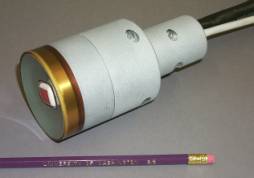
Integrated Transducer Probe (imaging array is inside the
white bordered area) The aluminum housing is watertight, and has been
designed to hold the imaging scanhead precisely. |
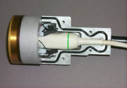
Final assembly included seating and
sealing the probe with rubber molding compound for a shock absorbing
fit. The 32 HIFU channels (64
signal and ground wires) are connected to a cable terminating inside one half
of the split housing. |
|
|
|
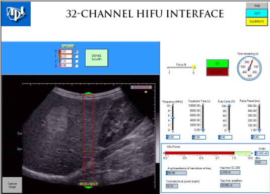
Four focal locations are
programmable in this panel, as well as various other treatment parameters
that define a HIFU dose (Frequency, Treatment time, Duty Cycle, Pulse Period,
HIFU Power). |
The HIFU transducer is a 32-element annular array made from a thin spherically shaped piezoceramic bowl that operates at a nominal frequency of 2 MHz. The ceramic is bonded to a mechanical matching layer and cut into 32 concentric annuli. Driving each individual ring with an independently programmed and amplified signal allows adjusting the location of the focal zone (hot spot) within a range of about 4 to 12 cm from the transducer along its axis (1-dimensional adjustment). As mentioned earlier, a central opening in the transducer permits mounting of a commercial medical imaging phased array scanhead (ATL P7-4) that is held precisely in place within the special housing illustrated above. This mechanical fixture ensures precise registration between the HIFU transducer axis and the image plane of the imaging probe. The multi-channel electronics system is capable of generating up to 1024 independent channels with programmable amplitude, phase and frequency signals. A large channel count is needed for more flexible control of the focal zone location in transverse as well as longitudinal position; this is possible using HIFU transducer arrays with many individual elements arranged in a 2-dimensional grid. To drive the annular array, we have built 32 dual-stage linear power amplifiers capable of about 40 W continuous duty each, and have combined the electronics in a wheeled rack for use in a research clinic.
|
|
|
|
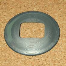
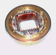
Photo of
the back of the spherical piezo element, cut into annuli and with a cutout
for the imaging probe. |
An image
of the back of transducer, after mounting and ready for electrical
connection. |
Researchers all over the world are developing HIFU technology and evaluating clinical protocols in animal and human trials. In China, several groups have treated thousands of patients for many types of cancer with very promising results. At the University of Washington, we are pursuing the development of new HIFU tools and techniques to stop bleeding (hemostasis) and to treat troublesome benign and malignant tumors. We are also developing novel imaging algorithms to greatly improve the ultrasonic detection and delineation of internal bleeding sites, and of currently undetectable newly forming cancerous tumors. In conclusion, while in many ways less sophisticated than the new molecular and genetic treatment approaches under development today, image-guided ultrasound therapy may also prove to be a revolutionary advance in surgical practice.