[ Lay Language Paper Index | Press Room ]
Azra Alizad, Alizad.Azra@mayo.edu,
F. G. Mitri, R. R. Kinnick, J. F. Greenleaf, and M. Fatemi
Mayo Clinic College of Medicine
Rochester, MN 55905
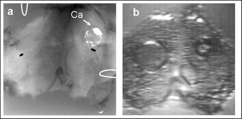
Popular version of paper 3pBB3, "Vibro-acoustography of the thyroid"
Presented Thursday afternoon, November 29, 2007 at 2:05 p.m.
154th ASA Meeting, New Orleans, LA
The incidence of thyroid cancer is increasing faster than any other tumor type at present. The reason for this growth is that we are getting better at detecting thyroid cancer, thanks to the increased use of ultrasound in clinical thyroid practice. It is a high sensitivity tool for detecting small thyroid nodules, including thyroid cancers.
However, 95% of these newly discovered thyroid nodules are benign. But determining the significance of solid thyroid masses is difficult using current imaging modalities. Fine needle aspiration or biopsy is the most accurate method of characterizing thyroid nodules. Development of an imaging method to more accurately determine malignancy or benignity could eliminate many biopsies of indeterminate nodules. Ultrasonography may be useful in distinguishing among cysts, cystic tumors, and solid tumors, but ultrasonography alone is not diagnostic.
Digital palpation, the examination of tissue through the use of touch, remains one of the simplest yet effective methods for detecting thyroid nodules. Thyroid tumors are often much harder than the normal tissue, and sensing stiff tissue can reveal early signs of a tumor. However, the sense of touch is not sensitive enough to detect small or very deep lesions.
Significant effort has been invested in developing improved imaging techniques, especially those that provide palpation-like information. Vibro-acoustography (VA) is an innovative, non-invasive imaging method that is sensitive to tissue stiffness. Typical VA images have high resolution and high contrast. Both soft tissue and microcalcifications can be imaged with VA, making this method is an ideal tool for tissue imaging. It is currently in the experimental stage and cannot be used for clinical practice in the present form.
This paper describes recent results on imaging human thyroids with vibro-acoustography. Experiments were conducted on excised human thyroids form autopsy. Vibro-acoustography displayed calcifications, anatomical details, and some nodules where they existed. Furthermore, vibro-acoustography images displayed tissue structures with high contrast and free from snowy pattern seen in ultrasound. Vibro-acoustography may be a suitable technique for thyroid imaging, though more data from a larger population are needed to establish the results in a statistical sense.