Investigation of an Ultrasound Imaging Technique to Target Kidney Stones
Anup Shah - anupshah@u.washington.edu
Dept. of Urology, Univ. of Washington School of Medicine
1959 NE Pacific St., Box 356510, Seattle, WA 98195
Marla Paun, John Kucewicz, Oleg A. Sapozhnikov
Center for Industrial and Medical Ultrasound, Applied Physics Lab.Univ. of Washington
Seattle, WA 98105
Manjiri Dighe
Dept. of Radiology, Univ. of Washington School of Medicine
Seattle, WA 98195
Hunter A. McKay
The Polyclinic
Seattle, WA 98122
Mathew D. Sorensen
Dept. of Urology, Univ. of Washington School of Medicine
Seattle, WA 98195
Michael R. Bailey - bailey@apl.washington.edu
Lawrence A. Crum
Center for Industrial and Medical Ultrasound, Applied Physics Lab., Univ. of Washington
Seattle, WA 98105
“Twinkling artifact” on Doppler ultrasound imaging is known and has been proposed before as a way to detect kidney stones. A Doppler mode exists on clinical ultrasound to detect motion, particularly blood flow, and display the moving blood as red or blue on the screen; however, for some unknown reason, when a stationary kidney stone is imaged in Doppler mode, the stone is displayed as a rainbow of colors, which makes the stone readily apparent. Something about the presence of the stone tricks the machine into displaying the color, which is an “artifact” because the color no longer represents true motion. Because it is a trick on the machine, an artifact can be intermittent and unreliable. The unreliability is exacerbated because each model of ultrasound imager has different and proprietary technology. Our talk focuses on how we are going about understanding the artifact and making it into a useful tool in detecting and treating kidney stones.
|
|
|
|
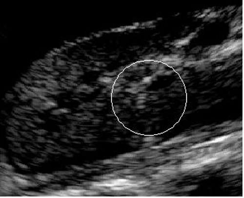
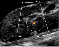
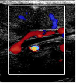
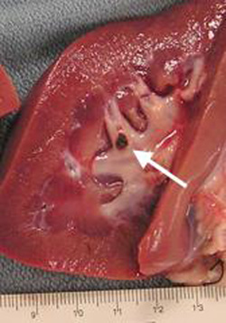
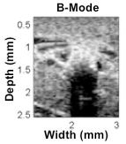
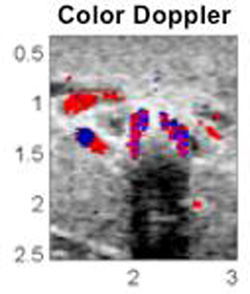
Figure 1. Illustration of the twinkling artifact. Shown in A and B are ultrasound images of a 2 mm stone inserted into an in vivo kidney. In A, the scan is only B-mode; in B, color Doppler is engaged. Note that the twinkling artifact shows a well defined stone (circled) while the B-mode image shows no obvious indication of a stone. Shown in C is a stone placed near a blood vessel and imaged in vivo; note that it is easy to distinguish between the stone exhibiting the twinkling artifact and regular blood flow. Frame D shows a photograph of the stone placed in the kidney. |
|||
Kidney stones afflict 13% of men and 7% of women in the U.S., and these numbers are rising. The prevalence of kidney stones varies dependent on race, sex, age, and geographic location; however, stone formers can be any age and many develop multiple stones at a time. Some stones pass spontaneously; however, those that don’t account for over 2 million outpatient treatments and 1% of all hospitalizations annually in the U.S. Total treatment cost in 2000, 8 years ago, was calculated to be $2.1B, which was 50% higher than 6 years earlier (1994).
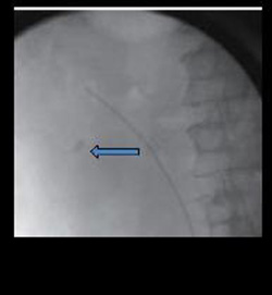 Figure 2. Fluoroscopy image of a kidney stone.
We see at least three applications of twinkling artifact to kidney stones. First stones are usually in the U.S. diagnosed with spiral CT imaging, which is not done in the doctor’s office and exposes the patient to ionizing radiation. Conceivably our ultrasound technique would allow an immediate localization of the stone in the office and space the patient the radiation exposure. Second, most stones in the U.S. are treated by lithotripsy, where shock waves are sent into the patient’s body to break stones. Most often x-ray fluoroscopy, which generally is not as good as spiral CT, is used to find the stone to target the treatment. Figure 2 shows a fluoroscopy image of a stone. These images are not always clear and sometimes the lithotripsy has to be done based on a best guess as to the location the stone. The stone in Fig. 2 is not as clearly identifiable as the one in Fig. 1B, so potentially twinkling could provide better targeting without the X-ray radiation. Third, the stone moves as the patient breathes during lithotripsy treatment, which means that about half the shock waves miss the stone and impact only kidney tissue. Lithotripsy is known to have a side effect of tissue injury, and the fewer shock waves used, the less injury. Twinkling on ultrasound is a sensitive and real-time stone detector that could be used to ensure shock waves are only triggered when the stone is in the focus.
Although the applications are clear, the mechanism that causes twinkling is a complex mixture of factors. The ultrasound machine does produce tiny motion of the stone and receives from the stone an echo that is generally stronger than that from tissue and contains reverberations from within the stone. These extra signals appear as if structures within the volume of the stone are moving in and out of the image. The confusion is further compounded by processing within the machine, which essentially amplifies the extra signals and variation in the collection of sequences of images. Our approach has been to use numerical modeling of the echoes and reverberations, Fig.3, and compare that to the raw data collected by ultrasound machines for stones in water, tissue, and patients. We then create our own images using specific algorithms that mimic the proprietary processing in the imaging systems. We can generally recreate what is shown on the imagers and detect patterns that are used to specifically image just stones not motion, as shown in Fig. 4. In our experience, the artifact has reliably revealed the stone in 100% of the animal studies and in an initial handful of human studies.
|
|
|
|
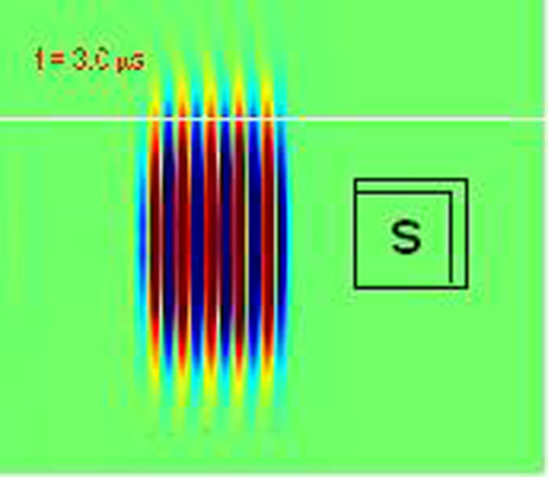
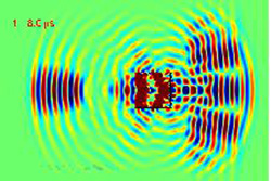
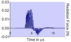
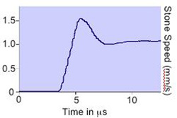
Figure 3. Modeling results that suggest acoustic radiation force as a possible reason of the “twinkling” artifact. (A) Acoustic pressure distribution when ultrasound diagnostic pulse, being directed to a kidney stone (s), is scattered (B). The scattering gives rise to an oscillatory radiation force imparted on the stone (C). The stone is thus pushed and starts to move. The corresponding speed (D) is high enough to create a noticeable Doppler signal for small stones, <1 mm. Larger stones can be also pushed in the transverse direction, which may result in changing the ultrasound speckle pattern and also contribute to the artifact. |
|||
|
|
|
|
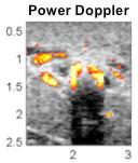
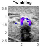
Figure 4. Images generated on post-processing of raw data from the ultrasound imager: A) B-mode shows shadowing but little of the stone, B) and C) normal Doppler processing shows the artifact on the stone as well as blood flow, and D) new processing for the distinct signal of twinkling adds color only to the stone. |
|||
WATCH: Video of kidney stone twinkling