![]()
![]()
![]()
Charles Caskey
- cfcaskey@ucdavis.edu
Mario Hlawitschka
Shengping Qin
Katherine Ferrara
Dept. of Biomedical Eng., University of California at Davis
451 Health Sci. Dr., Davis, CA 95616
Popular version of paper 4aBB2
Presented Thursday morning, November 18, 2010
2nd Pan-American/Iberian Meeting on Acoustics, Cancun, Mexico
Although ultrasound imaging is probably best known for its diagnostic capabilities in echocardiography and prenatal care, ultrasonic waves can also be used for therapy by focusing an acoustic beam through the skin and into the body. An exciting application for focused ultrasound is localized delivery of drugs within the body.
Because a drug is only effective after reaching its intended target in the body, the method of ingestion and route the drug takes to its target is important. Many drugs can be ingested by simply taking a pill that is absorbed through the stomach; however, this is not possible with some drugs, such as toxic cancer-fighting chemotherapeutics, which kill any cells they come into contact with—regardless of whether the cell is cancerous or part of the body. To address the problem of transporting a drug to a targeted location within the body, researchers will often encapsulate drugs in a shell. The drug is inactive until the shell is broken or ingested by targeted cells. These shells, often called drug delivery vehicles, are used clinically and have undergone many developments in recent years. The development discussed here is the guidance of drug delivery vehicles that can be activated by external forces, such as heat from a therapeutic ultrasonic beam.
Focused ultrasound transducers generate acoustic waves for drug delivery by concentrating acoustic energy generated by the transducer into a small focal region (typically about the size of a grain of rice) where the energy is absorbed and converted into heat. Although the location and size of the transducer’s focal region is well defined, guidance of the small focus within the body is difficult because the body is opaque. Furthermore, the grain-sized focal zone is often much smaller than the region targeted for heating, so the beam must be moved around in order to treat the entire target region. Therefore, acoustic therapy without guidance is analogous to being lost without a map.
Fortunately, three-dimensional imaging systems, such as x-ray computed tomography, are capable of generating excellent maps of structures within the body that can be useful for guiding acoustic therapy. However, many a lost individual can attest that maps are only useful if you can find your current location on the map. Fortunately for these individuals, there are now satellites in space that enable anyone owning a GPS device to see their current location and orientation on a digital map. The solution for guiding acoustic therapy based on a computed tomography map is analogous in that the computed tomography images provide a map of the body, while a tracked sensor attached to the therapeutic transducer plays the role of the positioning system. Whereas GPS allows a device to know its location relative to satellites orbiting the earth, the positioning system used for therapeutic ultrasound functions by tracking a sensor relative to a magnetic field emitter or infrared camera that is placed near the object being imaged (see Figure 1). The sensor is attached to the therapeutic ultrasound transducer such that the transducer location can be determined relative to the diseased region. The tracking technology is similar to that used by the Nintendo Wii controllers, except that the transducer sensors for therapeutic ultrasound must be much more accurate to ensure high quality therapeutic guidance.
By combining tracked therapeutic ultrasound with computed tomography, one can know with very high accuracy exactly where the transducer focal region is located inside the body. Acoustic therapy can then be planned in a similar way to planning a route on a navigational GPS device. Instead of generating a list of directions based on a map of roads, the guided therapy system uses the computed tomography map to characterize tissues as fat, bone, or protein (see Figure 2). These tissues are characterized because each absorbs ultrasound differently and therefore undergoes heating at different rates. Through this characterization, our tracked imaging system can determine the ultrasound properties required for treatment so the ultrasound beam can be controlled to successfully activate drugs for tumor therapy.
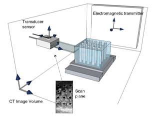
Figure 1. Coordinate systems utilized in joining ultrasound and CT image volumes. The transducer sensor location is detected relative to the electromagnetic transmitter, so that the ultrasound scan plane and focal point can be found in the computed tomography (CT) image volume.
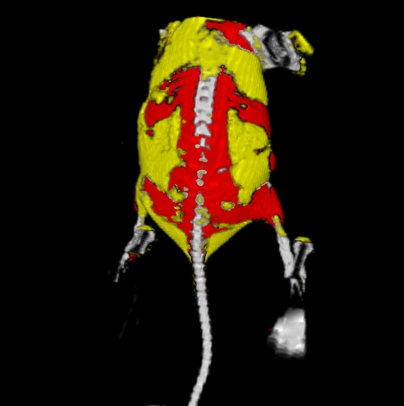
Figure 2. A computed tomography map of a mouse with tumors in the hind limps. The fatty regions, bone, and protein-rich regions of the mouse are represented by the yellow, white, and red, respectively.