Dimitri M. Donskoy - ddonskoy@stevens-tech.edu
Davidson Laboratory
Stevens Institute of Technology
Hoboken, NJ 07030
Popular version of paper 4aBV13
Presented Thursday morning, June 19, 1997
133rd ASA Meeting, State College, PA
Embargoed until June 19 1997
Over 20 million American women suffer from osteoporosis, according to the American Academy of Orthopaedic Surgeons [1]. Osteoporosis, " silent crippler ", is a bone disorder resulting in bone mass reduction due to hormonal changes associated with aging. Bones become fragile and more likely to break. Osteoporosis costs the nation over $10 billion annually more than congestive heart failure or asthma [2]. The World Health Organization recently placed osteoporosis on the priority list of the non-transmissible diseases for which preventive programs have to be implemented throughout the world.
Diagnosis of osteoporosis is based on bone density measurement. Photon and X-ray absorptiometry and X-ray quantitative computed tomography are routinely used in clinical practice for these measurements [3]. However these techniques have limited applicability because of the expensive and bulky equipment and procedure, and the potential risk from radiation.
Studies of the application of acoustic energy for non-invasive skeletal diagnosis have shown its feasibility and demonstrated the advantages of utilizing acoustic (ultrasonic and subsonic) techniques for bone mass and strength measurements. Unlike conventional radiological techniques, acoustic techniques emit no radiation, are cost effective, and utilize equipment which is portable and easy to operate.
The ultrasonic technique uses correlation between measured speed and attenuation of high frequency sound waves (ultrasound with frequencies tens to hundreds of kHz) propagating through bone tissue and the density of the tissue. Although a significant number of ultrasonic tests have been performed, this technique is not widely used as a bone diagnostic tool for clinical application due to some difficulties in the interpretation of the measurements. Thus, the speed and attenuation of ultrasonic waves depend on density as well as on certain other properties of bone. According to the study [4], only 53% of the attenuation value and 44% of the speed value can be accounted for by density.
Subsonic techniques utilize low frequency acoustic energy (hundreds of Hz, or oscillations per second) often called vibration. Subsonic methods, known as impedance and resonance methods, involve excitation and measurements of flexural mode of vibration of long bone and show some potential applicability for osteoporosis diagnosis. However, the interpretation of the subsonic measurement is also a difficult task and to a great extent, depends upon a corresponding mathematical model of the tested object. The influence of surrounding soft tissue (its mass and damping effect) creates additional difficulties in the interpretation and use of these methods.
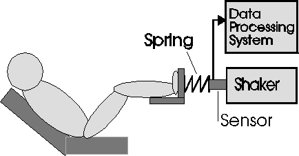
The proposed infrasound method is sufficiently different from the existing methods. Instead of measuring flexural resonances, which involve both the mass and flexibility of a bone and the surrounding soft tissue, the method based on measurements of a " rigid body " resonance of a long bone, such as tibia or ulna, with the use of an artificial spring with known stiffness. This approach greatly simplifies the interpretation of the measurements and is more accurate.
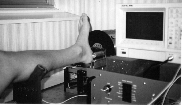
The method employs very low frequency (below the audible range) longitudinal vibration excitation of human long bone through an artificial spring as illustrated in Fig.1. The spring is much softer than the bone and its joints, but stiffer than surrounding soft tissue. Because of this, the flexibility of the bone and joints as well as mass of the surrounding soft tissue have no effect on longitudinal oscillation of the spring-bone system, where bone contribution is purely inertial. Thus, from mechanical point of view, the bone can be considered as a rigid mass. The spring-mass system exhibits resonance behavior (amplification of the oscillation on the particular frequency, called the resonance frequency). The resonance frequency depends on the stiffness of the spring and the attached mass. Therefore, by measuring the frequency (the spring stiffness is known) the overall mass of bone can be determined. An important feature of this approach is that the resonance frequency can be shifted into very low frequency range (infrasound range) by proper choice of the spring stiffness. This eliminates the damping effect of soft tissue raising the sharpness of the resonance and, hence, increasing the accuracy of the measurements.
Numerical analysis of the developed model has shown that the mass of bone and its loss can be accurately measured and these measurements do not depend on variation in bone flexibility and soft tissue parameters. An experimental setup (Fig.2) was arranged and in-vivo measurements were conducted and proved the basic concept of the proposed method.