Xiaoming Zhang - Zhang.Xiaoming@mayo.edu Popular version of paper 3aBB9 and
3aBB8 Vessels are important parts of our
body. Their function is critical to our well-being. For years, doctors have
tried various tools to take pictures of arteries that would help them in diagnosing
or preventing diseases. This has not been an easy task, however, and sometimes
they have to use imaging methods that may have some harmful side effects. It
has always been a challenge to take clear pictures of vessel without the negative
side effects. Here, we suggest a new way that may help us to achieve this goal.
Vessels in our body are narrow tubes,
with many branches along the way. Each segment may be thought of as a string,
much like a piano string, extending from one point to another. Hammering on
a piano string helps a tuner to "visualize" the string and find out if the string
is in a good condition or not. We use the same concept, but with more sophistication,
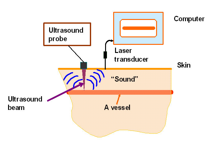
in our new imaging method. In our method, we tap a vessel to make it vibrate
at its natural tone, and we record the sound produce by the "singing" vessel.
As we move the tapping point across the area around the vessel, we record the
sound and map point-by-point to make an image. The sound would be strong only
if we are tapping on the vessel. The result is a "picture" of the vessel. This
picture actually shows the vibration or sound of the vessel, so we may call
it a "sound" image. If there is a change in vessel properties, for example due
to a disease, the change will effect the natural tone of the vessel and therefore
would be visible in this "sound" image.
In practice, the tapping is done
with a specially designed ultrasound probe and the sound is recorded by a sensitive
laser system that detects vessel vibrations from outside. This way we do not
need to have direct access to the vessel. The method is considered safe because
we do not use harmful radiations. Our initial experiments on objects resembling
human vessels have shown promising results.
Mostafa Fatemi
James F. Greenleaf
Ultrasound Research Laboratory
Mayo Clinic
Rochester, MN 55905
Presented Wednesday morning, April 30, 2003
145th ASA Meeting, Nashville, TN
