146th ASA Meeting, Austin, TX
[ Lay Language Paper Index | Press Room ]
Pumping Up the Ear
David C. Mountain, Ph.D. - dcm@bu.edu
Hearing Research Center
Boston University
Boston, MA
K. Domenica Karavitaki, Ph.D.
Harvard Medical School
Department of Neurobiology
Boston, MA
Popular version of paper 4pABa1
Presented Thursday afternoon, November 13, 2003
146th ASA Meeting, Austin, TX
New images of movements inside the cochlea (the auditory portion of the
inner ear) suggest that the incredible sensitivity of mammalian hearing
may be the result of cells that act as electromechanical fluid pumps.
These cells, called outer hair cells, are arranged in three rows within
the organ of Corti (Figure 1a) and are known to exhibit voltage changes
in response to acoustic stimulation [1] and to exhibit length changes
in response to changes in membrane voltage [2]. Scientists have
been puzzled, however, by the question of how these responses could increase
our ability to hear faint sounds. The new images show that when
the outer hair cells contract, they push fluid back and forth through
a tiny channel in the sensory organ called the tunnel of Corti (Figure
1a). Theoretical calculations show that, if this fluid flow is properly
synchronized with the sound-induced motions in the cochlea, then hearing
sensitivity can be increased 100-fold.

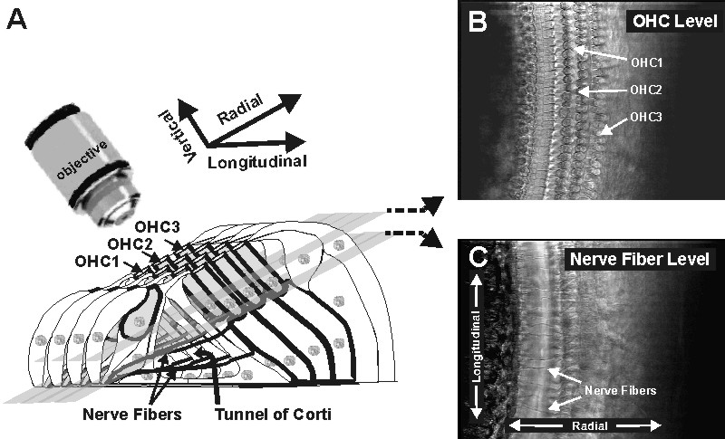
Figure 1. Imaging the organ of Corti. A) A diagram of the organ of Corti showing the position of the microcope objective. The gray planes show the imaging levels used for panels B) and C). B) Image from the level of the outer hair cells showing the three rows (OHC1, OHC2, and OHC3). C) Image from the level of the nerve fibers showing the fibers crossing the tunnel of Corti.The mammalian cochlea consists of a fluid-filled tube that is coiled like a snail shell, hence the name cochlea (the Latin word for snail). This tube is divided along its length by a membrane, the basilar membrane, and it is upon this membrane that the organ of Corti is found. When the cochlea is excited by sound, vibrations travel down the basilar membrane and stimulate the hair cells. The motions involved are extremely small, typically less than 1/1000th of the length of the cells.
To image the extremely small but very rapid vibrations present in the cochlea, we used stroboscopic illumination flashing at rates up to 10,000 times a second to “freeze” the apparent motion. Images were then captured with a computer-controlled video system mounted on a microscope [3]. The outer hair cells were electrically stimulated in order to change their membrane voltages at frequencies that spanned much of the normal hearing range. By changing the time delay between the strobe flashes and the electrical stimulus, it was possible to capture a series of images that could then be animated on a computer display. The animation procedure produces a slow-motion video of vibrations that were in fact taking place at a rate of 100s or even 1,000s of times per second. Computer vision techniques were then used to quantify the movements that could be as small as 10 nanometers, or about 1/1000th of the length of a hair cell.
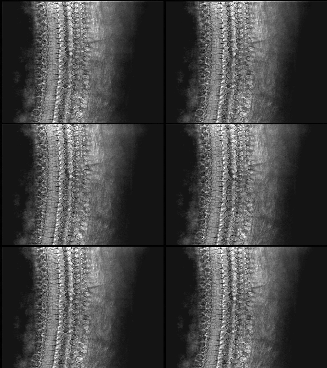
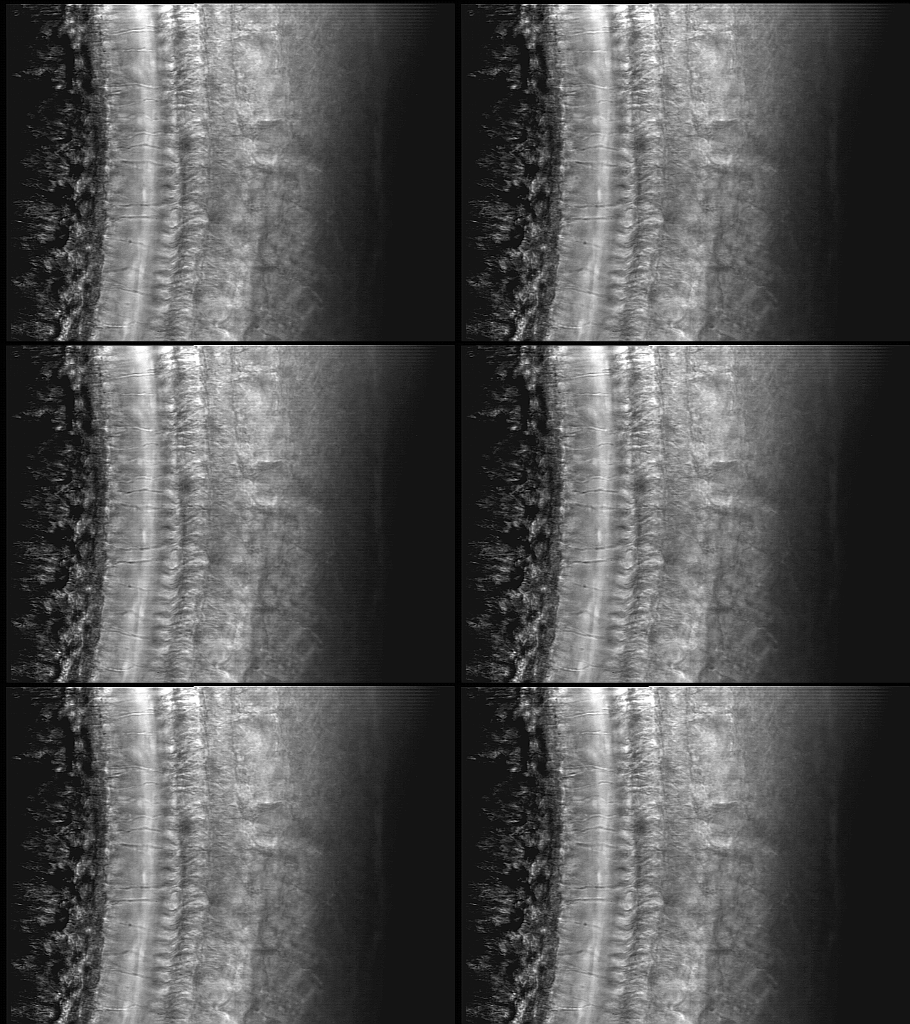
When we focused at the level of the outer hair cells (Figure 1b), we observed that first and third rows of outer hair cells moved apart when the cells contracted (Movie 1). This type of motion suggests that the hair cell contractions are causing a pressure increase within the organ of Corti that causes the cells to spread apart in the radial dimension. When we focused deeper into the organ (Figure 1c), we observed longitudinal displacements of nerve fibers that traverse through the tunnel of Corti (Movie 2). These displacements were synchronized with the outer hair cell contractions and appeared to be due to the longitudinal movement of fluid within the tunnel. We find that this motion of the fibers can be observed in regions several millimeters away from the region where the outer hair cells are contracting. We hypothesize that, when the outer hair cells contract, they squeeze the organ of Corti and pump fluid into the tunnel.

Movie 1. Outer hair cells are contracting in response to electrical stimulation

Movie 2. Nerve fibers are moving in response to electrically evoked outer hair cell contractions
Among all the vertebrate hearing organs, only mammals have a tunnel of Corti and only mammalian ears have hair cells that change their length in response to membrane voltage. Our ability to visualize the very small but very fast movements within the inner ear has given us the opportunity to shed some light on how these features contribute to our ability to hear faint sounds. The novel aspect of our findings is that, as a result of OHC contractions, fluid flows in the tunnel of Corti. These experimental findings suggest that fluid flow may be critical for our amazing hearing sensitivity.
REFERENCES
1. Dallos P, Santos-Sacchi J, Flock A. (1982) Intracellular recordings from cochlear outer hair cells.
Science. 218:582-4.
2. Brownell WE, Bader CR, Bertrand D, de Ribaupierre Y. (1985) Evoked mechanical responses of isolated cochlear outer hair cells. Science. 227:194-6.
3. Karavitaki, K.D., Mountain, D.C. and Cody, A.R. (1997). Electrically-evoked micromechanical movements from the apical turn of the gerbil cochlea. In: Diversity in Auditory Mechanics ER Lewis, GR Long, RF Lyon, PM Narins, CR Steele, and E Hecht Poinar eds, World Scientific Publishing, pp. 392-398
4. Hubbard, A.E., Shatz, L., Yang, Z and Mountain, D.C. (2000) Multimode cochlear models. In: Symposium on Recent Developments in Auditory Mechanics, H. Wada, T. Takasaka, K. Ikeda, K. Ohyama, T. Koike eds. World Scientific Publishing, Singapore, pp. 167-173.