ASA/CAA '05 Meeting, Vancouver, BC


The Healing of Bone - A New Field for Shock Waves?
Joerg Hausdorf- joerg.hausdorf@med.uni-muenchen.de
Volkmar Jansson
Markus Maier
Michael Delius
Ludwig-Maximilians-Univ.
Munich, Marchioninistr. 15
81377 Munich, Germany
Popular version of paper 1aBB3
Presented Monday morning, May 16, 2005
Joint ASA/CAA Meeting, Vancouver, BC
Extracorporal shock waves are transient pressure disturbances
that propagate rapidly in three-dimensional space. For more than 20 years, urologists
have used devices which are able to focus those shock waves onto a small target
to treat kidney stones. Those waves migrate without damage through skin and
soft tissue and set their energy free on the borderline of a tissue with a far
different density like calcified kidney stones ... or bone.
For patients who underwent multiple lithotripsies (shock wave treatment of kidney
stones), physicians found new growth of bone in the iliac crest (the major part
of the pelvic bone). This phenomenon was recognized as a reaction to the shock
waves and researchers began to study those bony reactions. First it was thought
that high energy was needed to somehow break the bone and then cause it to heal.
But recent studies showed that even lower energy levels, which did not show
any destructions of the bone, were able to enhance bone healing.
In our group, which has been working in shock wave research for more than 25
years, we were able to show that at the scale of the cell, our lowest level
of biological organization, there is a reaction to shock waves. We applied shock
waves to fibroblasts (cells from soft tissue like tendons, ligaments or muscles)
and to osteoblasts (cells which form the bone). One day after the treatment
we found an increase of a very important bone growth factor (bFGF) identically
in both cell types. So for the first time it could be shown that shock waves
enhance the production of this bone growth factor in intact human cells (Fig.
1).

Fig. 1: bFGF production in osteoblasts
What may be the use of these findings? In orthopedics we know some diseases or posttraumatic conditions where we are still looking for a successful therapy with low complications and low costs. This concerns the necrosis (death) of the femoral head (the ball-and-socket joint of the hip), where bony substance dies, the highly loaded hip joint gets damaged, patients have lots of pain and end up with a hip replacement. It also concerns all fields of non-union of bones, in which physicians must come up with. procedures to take care of fractures that have not fully healed. We already know that the right amount of growth factors would help in these conditions, but the question is how to carry the substances to the right area, how to create these substances without the risk of infection, allergy or cancer and not at least how to keep the costs down.
So for the femoral head necrosis we wanted to know whether the
shock waves are really able to migrate deeply enough into the bone to reach
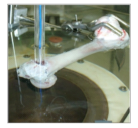
the necrotic area. Therefore we measured the pressure inside the femoral head
of cadaver pig bones while shock wave application. And indeed we found even
10 mm beneath the surface still 50 % of the shock wave pressure.

Fig. 2: Measuring pressure during shock wave application to pig bones
This leads us to continue our research in this field because the extracorporal shock wave therapy would be a noninvasive method with low complication rate to heal pathologic bone conditions with body's own substances.