ASA/CAA '05 Meeting, Vancouver, BC


Helping Doctors Interpret the Sound of Blood:
Using a Multimode Sonic and Ultrasonic Imaging System
Helping Doctors Interpret the Sound of Blood:
Using a Multimode Sonic and Ultrasonic Imaging System
Thomas Royston - troyston@uic.edu
T. Spohnholtza), T. J. Roystonaa), Y. Yaziciolgua),
B. A. Martina), F. Lotha), H. Bassiounyb)
a)University of Illinois at Chicago
Chicago, IL 60607
b) University of Chicago
Chicago, IL 60612
Popular version of paper 5aBBb8
Presented Friday morning, May 20, 2005
Joint ASA/CAA Meeting, Vancouver, BC
Measurement of naturally occurring sounds in the body, such as those caused
by breathing, by the heart pumping and by blood flowing, can augment conventional
medical imaging technology by providing unique information about system function.
This paper describes how a device to precisely measure these sounds has been
inexpensively integrated into a portable ultrasonic imaging platform to increase
what can be learned about how blood is flowing in critical vessels in the body.
Atherosclerosis is one of many vascular diseases that result in a narrowing
or blocking of blood vessels. A narrowing or constriction of a vein also often
happens due to vein wall thickening just downstream of arteriovenous grafts,
which are used in many patients with advanced diabetes to aid in dialysis. This
wall thickening may be in response to irregular blood flow patterns that occur
downstream of the constructed graft.
Current methods to detect such constrictions in blood vessels and identify associated diseases include angiography and ultrasonic imaging. Angiography is an invasive procedure in which a catheter is surgically inserted directly into an artery and releases dye. Blood flow is then tracked by visualizing the dye, generally by exposing the patient to X-rays, to find areas of reduced blood flow.
Ultrasonic imaging uses very high-frequency sound waves to construct images of the blood vessel geometry that can be used to estimate size and shape. Color Doppler ultrasound also provides an estimate of the blood velocity in a region, which may help identify a constricted zone. Ultrasonic imaging is non-invasive; so no surgery or exposure to ionizing radiation is required. But, it has limitations. In the newly developed multimode technique described here, the advantages of ultrasound, its noninvasiveness and high resolution imaging of geometry, are combined with the capability to detect low-intensity audible (low frequency) sounds that are associated with vessel constrictions and other factors that cause irregular blood flow.
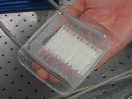
The presence of an obstruction in the vessel disturbs the flow of blood. This disturbance in turn generates unique sounds that travel to the outer surface of the skin. These sounds can be detected by sensitive acoustic sensors arranged in an array, shown in Figure 1. Through specialized data processing techniques, the signals acquired from the sensor array can be used to estimate the 3-dimensional sound field in the vicinity of the array and the location of specific sound sources.
The technique is further enhanced by developing the acoustic sensor array to be both flexible and transparent to ultrasonic waves. Flexibility allows the pad to conform to curved surfaces of the body such as arms or legs. Though not transparent to visible light, the pad is invisible to ultrasound, which allows a commercially available ultrasound system to acquire data directly through the acoustic pad while the pad simultaneously acquires acoustic (sonic) data.
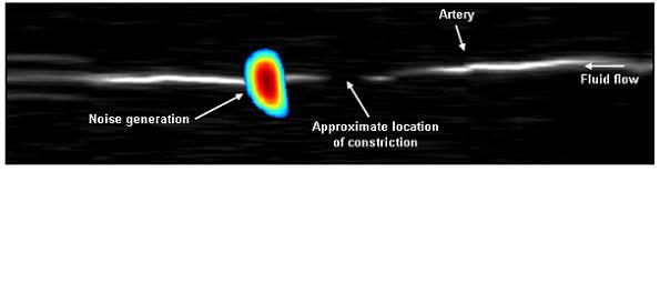
The calculated sound field is then combined with the geometry obtained from ultrasonic imaging to produce a composite image. Figure 2 shows the multimode image of a simulated blood vessel in the lab embedded in silicon, a material similar to soft human tissue, with fluid flowing through the vessel from right to left. The vessel's geometry as determined by ultrasound is represented by the grayscale portion of the image. The vessel has a small constriction present at the center of the image to simulate a moderate blockage and is at the point where the image of the vessel fades slightly. The acoustic field information obtained from the acoustic sensor array is displayed in color and shows a region of noise. This occurs just downstream of the blockage and clearly indicates the presence and approximate location of the blockage, if the direction of blood flow is known, say for example from the Doppler mode of conventional ultrasound.
This bi-modal approach to assessing vascular obstructions and possibly even predicting future ones by assessing irregular blood flow patterns is synergistically better than the individual methods. While the ultrasound image will usually indicate the presence of a constriction and give some information about its shape and approximate the blood velocity in a region using the Doppler mode, the sonic image confirms the presence of the constriction and gives us unique information about how the blood flow is affected by this constriction. Additionally, it may also predict the formation of a future constriction that results from an irregular blood flow pattern, which may occur, for example, downstream of a vascular graft. The sonic array measurement can tell us whether the flow is still relatively smooth and laminar or has become turbulent. This type of information may be useful in assessing the relative danger of the constriction and answer such questions as, how severe is the constriction and what types of forces is the blood vessel wall being exposed to from the flow that may cause the vessel wall to remodel itself, thicken or possibly fail in the future. By non-invasively and simultaneously acquiring both ultrasonic and sonic data, and combining them in a single image, a medical professional may more readily determine the best treatment option. [Research support: NIH EB002511 and HL55296, and Whitaker Foundation BME RG 01-0198].