![]()
![]()
![]()
Iulian Voicu
Jean-Marc Girault - Jean-marc.girault@univ-tours.fr
Morgan Fournier-Massignan
Franck Perrotin
Denis Kouamé
UMRS “Brain and Imaging” INSERM U930, CNRS ERL3106
François Rabelais University, Tours, France
33 2 47 36 60 55
Popular version of paper 2aBB12
Presented Tuesday Morning, November 16
2nd Pan-American/Iberian Meeting on Acoustics, Cancun, Mexico
Introduction
In utero, monitoring of fetal well-being or suffering is today an open challenge, due to the high number of clinical parameters to be considered. An automatic monitoring of fetal activity, dedicated to quantifying fetal wellbeing, becomes necessary. For this purpose and to supply an alternative for the Manning test, we used an ultrasound multi-transducer multi-gate Doppler system. One important issue (and first step in our investigation) is the accurate estimation of fetal heart rate (FHR). An estimation of the FHR is obtained by evaluating the autocorrelation function of the Doppler signals for ill and healthy foetuses. By modifying the existing method (autocorrelation method), we reduce to 5% the probability of non-detection of the fetal heart rate. These results are really encouraging, and they enable us to plan the use of automatic classification techniques in order to discriminate between healthy and suffering foetuses.
Materials
• 3 groups of 4 sensors, each sensor with 5 exploring depths
adjustable between 1.88 cm up to 15cm;
• Emission Frequency: 2.25 MHz;
• Acoustic Driving Power: 1mW/cm2;
• Pulse Repetition Frequency: 1kHz.
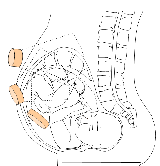
Figure 1. Positioning of the multi-sensors pulsed Doppler system.
Methods
Assessing the fetal heart rate consists of evaluating the envelope of a Doppler signal from which the short-time autocorrelation function is estimated. This estimation does not have to be corrupted by non desired signals such as mother’s heart rate, mother’s moving and pseudo breathing.
We claimed that the performances of the existing fetal heart rate can be largely improved by assessing the cardiac rhythm from directional Doppler signals rather than from the envelope Doppler signal. This hypothesis was based on the fact that i) the fetal heart rate is based on the estimation of the duration between local maximal peaks and ii) the Doppler envelope signal is too complex due to the presence of lot of peaks.
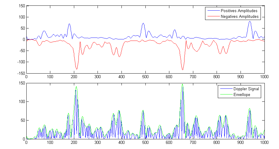
Figure 2. Directional and envelope Doppler signals
Unlike standard methods based on the estimation of the fetal heart rate (FHR) from the envelope signal (see Fig. 3a), we propose to estimate the fetal heart rate from directional signals (see Fig. 3b) (positive amplitude and negative amplitude).
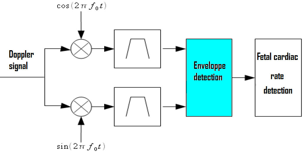
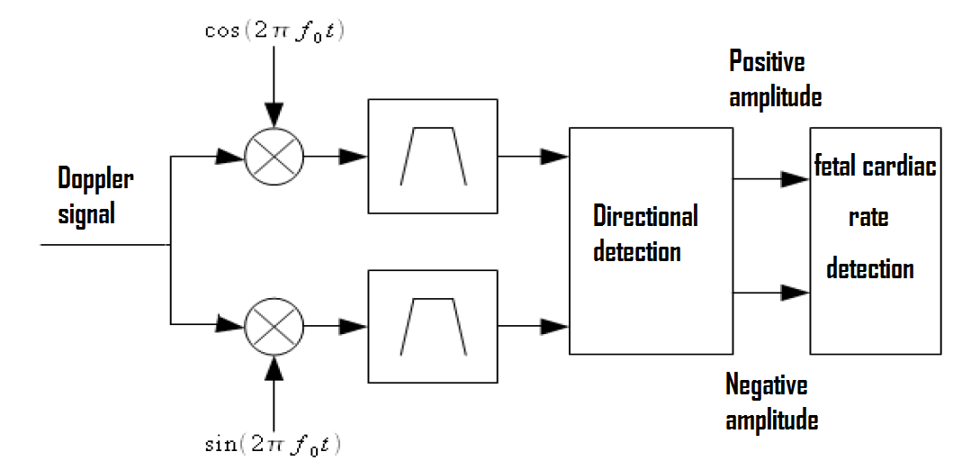
Figure 3. Functional schemes of the FHR envelope detector and the FHR directional detector.
Results and discussion
Using an analyzing window of 2.048 seconds, we show in Fig. 4 that the proposed
method gives FHR very close to the FHR given by the reference system Sonicaid. FHR obtained by the detector based on the
envelope Doppler signal shows some spurious peaks. These spurious peaks were
not observed for the reference detector and our new detector.
The performance of the statistical analysis of our new detector shows that the
false alarm rate can be reduced by a factor of 15% compared to performances of
the detector based on the envelope Doppler signal. These results seem to
corroborate our hypothesis based on the too complex nature of the envelope
Doppler signal.
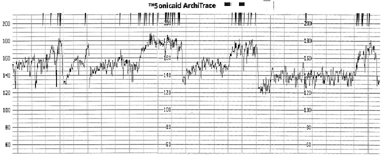
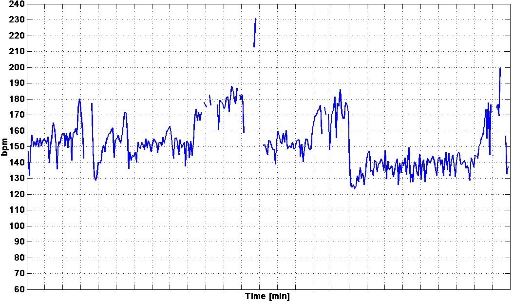
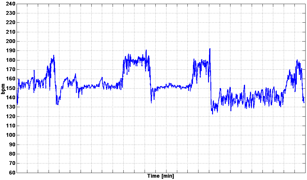
Figure 4. Fetal heart rate. a) Reference estimation of the FHR by the Sonicaid system. b) FHR estimation based on the envelope Doppler signal. c) FHR rate estimation based on the directional signals.
Conclusion
The auto-correlation is the method which gives the best trade-off in terms of probability of detection and number of operations. The size of the analyzing window and the overlapping step are very important. The good performance of our new detector is very encouraging and will permit us to propose a “fetal well-being electronic score” based on a reliable FHR estimation.