Figure 1. The relationship between high-speed video (all images) and stroboscopy (brighter images only) during vocal fold vibration.
Daryush Mehta – daryush.mehta@alum.mit.edu
Matías Zañartu
Thomas F. Quatieri
Dimitar D. Deliyski
Robert E. Hillman
Massachusetts General Hospital
Boston, MA 02114
Popular version of paper 3aSCa1
Presented Wednesday morning, November 2, 2011
162st ASA meeting, San Diego, Calif.
When we talk to each other, the delicate tissues of the voice box vibrate too fast for the naked eye to see or for standard video capture. Clever stroboscopic techniques allow voice specialists to observe many vocal characteristics, but irregular patterns of tissue vibration pose difficulties for such systems. Video 1 demonstrates the undesirable effects of a patient’s unstable voice on stroboscopic visualization.
Today, however, researchers and physicians are finally able to visualize and investigate irregular physiological behaviors, thanks to the development of a high-speed imaging system by an interdisciplinary research team at the Center for Laryngeal Surgery & Voice Rehabilitation at the Massachusetts General Hospital (MGH Voice Center). Team members include clinicians (Drs. Steven Zeitels and Hillman) and engineers/scientists (Drs. Mehta, Zañartu, Quatieri, and Deliyski), whose goal is to improve vocal health by understanding the movements of the vocal folds and their impact on voice quality. Using the same high-speed video technology that enables nature photographers to visualize the rapid beating of hummingbird wings, the Boston team has begun to observe and document details of vocal vibrations and voice acoustics that have never before been appreciated during clinical evaluations. The team will present their work at the annual meeting of the Acoustical Society of America in San Diego on Wednesday, November 2, 2011.
Explains research team member Daryush Mehta, PhD, “For the first time, voice scientists are able to investigate detailed relationships between vocal vibrations and sound qualities of the human voice. For example, we are just now discovering that certain asymmetric vibration patterns do not necessarily degrade one's voice acoustics as once thought.” This level of detail is possible due to recent technological advances in high-speed imaging of the larynx, including enhanced light sensitivity of digital camera sensors. It enables researchers to capture and analyze high-resolution video—exceeding 10,000 images per second—of the vocal folds that oscillate 100–1,000 times per second.
In one high-speed video setup, a rigid endoscope is passed through the mouth and positioned in the throat to provide magnified high-resolution color images of vocal fold tissue vibration. Video 2 shows high-speed video of the same patient above, capturing finer details of vocal fold motion. How much information do we miss with stroboscopic imaging? Figure 1 illustrates the additional level of detail obtained using high-speed video versus stroboscopy.
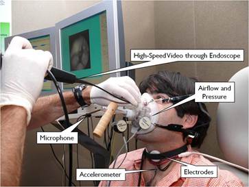
In the most comprehensive setup, researchers record multiple voice-related variables alongside monochromatic high-speed video that is captured with a flexible endoscope passed through the nose (see Figure 2). Additional variables include acoustic data and electrodes that relay information about when the vocal folds collide. Acceleration of the neck skin near the larynx arises from vocal fold vibrations and the airflow pulses the vibrations create. Finally, airflow and pressure variables document additional measures of vocal function.
With the comprehensive monitoring system, the research team hopes to improve clinical care of patients who have damaged or impaired voices. By helping pinpoint the deficits in vocal fold vibration, the high-speed video monitoring system can help identify anatomical areas that would optimally benefit from surgery or therapeutic treatment. In addition, new insight has already been gained regarding complex interactions between vocal fold vibration patterns, airflow, and laryngeal sound production.
The MGH work grew out of an urgent need to assess the true vibratory characteristics of vocal fold bio-implants that are currently under development at the MGH Voice Center. These bio-implants potentially aid in the restoration of soft tissue layers that are critical for humans to produce a healthy voice. “For detailed documentation of the restored tissue vibration, we turned to high-speed imaging technology,” Dr. Mehta explains. Because their system measures multiple variables, new levels of understanding—and possibly voice care—are now possible.
Media

Figure 1. The relationship between high-speed video (all images) and stroboscopy (brighter images only) during vocal fold vibration.

Figure 2. Research setup that simultaneously monitors high-speed video and multiple voice-related variables.
Video 1 -
Video 1. Stroboscopic recording of vocal fold vibration as a voice patient says “ahhh.” Note the inaccurate depiction of the patient’s irregular vibration pattern.
Video 2 -
Video 2. High-speed video (6000 images per second) of the same voice patient captures details of vocal fold motion not possible by stroboscopy. Contrast with Video 1.