Amit Katiyar
Mechanical Engineering
University of Delaware
Newark, DE 19701
Kausik Sarkar
Mechanical and Aerospace Engineering
George Washington University
Washington, DC
Email: Sarkar@gwu.edu
Secondary appointment:
Mechanical Engineering
University of Delaware
Newark, DE 19701
Krishna Sarker
Biological Sciences
University of Delaware
Newark, DE 19701
Popular version of paper 4pBA5
Presented Thursday afternoon, November 3, 2011
162nd ASA Meeting, San Diego, Calif.
Cancer is one of the leading causes of death among humans¾there were approximately 1.5 million new cancer cases and half a million deaths from cancer in 2010 costing $263.8 billion dollars for direct medical costs and indirect productivity cost (Cancer Facts and Figures, 2010). It is evident from the above statistics that the conventional treatments¾surgery, chemotherapy, radiotherapy, and hormone therapy are not enough. To effectively counter this epidemic, it is crucial that new strategies and tools are developed to fight this deadly disease. Here we propose to use the energy of ultrasound—sound wave pulsing at frequencies higher than the limits of human hearing—to cure cancer.

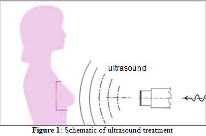
Ultrasound is best known for medical imaging; it is the safest and the most inexpensive means for peering inside the body to diagnose a disease. However, it also can deliver energy and mechanical stimulation remotely to targeted body parts. Mechanical stimulation can affect the biology and if used effectively can become a therapeutic tool. However, too strong an ultrasound application can also cause harm; strong bursts of ultrasound are used to break up kidney stones. Here, we propose application of a very weak ultrasound—LIPUS (Low intensity pulsed ultrasound)¾of less than 1Watt/cm2 power density for cancer treatment.
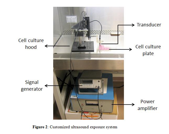
Cancer spreads by uncontrolled proliferation—increase of cell number. Controlling cell proliferation is critical to curing cancer. We show that even this weak ultrasound energy can significantly decrease cancer cell multiplication. We apply ultrasound to a human breast cancer line T47D, a popular target for cancer research. The ultrasound setup is shown in Figure 2.

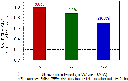
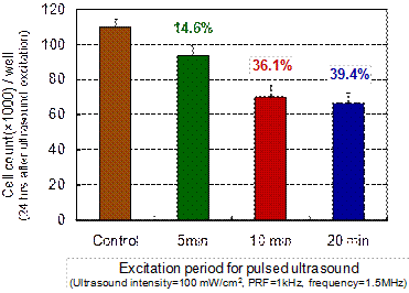
We grow the cells in the lab, put them in a cell culture plate, and stimulate them using a device called transducer in a sterile environment (cell culture hood). Rest of the setup shown in Figure 2 is used to drive the device. The cells are stimulated with ultrasound of increasing strength (intensity). Figure 3 shows that for the highest strength, we obtain a 30% decrease in cell multiplication. In Figure 4, total time of stimulation is varied to find that at least 10 minutes duration is needed to get this effect, and beyond that the beneficial effects do not increase.

Figure 3: Percent decrease in proliferation of ultrasound treated breast cancer cells at different ultrasound intensities.

Figure 4: Percent decrease in proliferation of ultrasound treated breast cancer cells at different ultrasound excitation durations.
In conclusion, this study demonstrates that low intensity pulsed ultrasound slows down proliferation of breast cancer cells. Cells sense the mechanical force and respond by changing their biochemical activities leading to this effect. The actual mechanism remains unknown, and needs to be explored in future research. Note that such low intensity pulsed ultrasound can heal fractures, and has been approved by the US Food and Drug Administration. There also the exact mechanism remains uncertain. The current experiment in a flat dish needs to be replicated in tissues and tumors in live animals before it will be ready for human trials. However, if successful it will be welcomed by millions whose lives have been devastated by the scourge of this fatal disease.