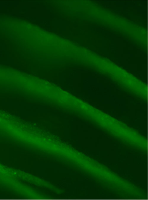
Figure 1. Banding of cells with ultrasound standing wave fields. The cell bands are spaced a distance of ~750 microns (Garvin et al. 2010).
Popular version of paper 4aBA5
Presented Thursday Morning, May 8, 2014
167th ASA Meeting, Providence
---------------------------------
There is a critical need for organs and tissues for patients in need of organ transplantation or tissue reconstruction. The field of tissue engineering focuses on developing technologies that enable the replacement or repair of diseased or injured tissues and organs. Recent advances in this field include the engineering of skin substitutes, cartilage replacements, and artificial bladders, all of which are relatively thin tissues. However, two critical challenges currently limiting the fabrication of larger more complex organs are 1) the need for patterning technologies that can reconstruct complex cell and tissue organizations, and 2) the need for a vasculature system in the artificial tissue to provide oxygen and nutrients throughout the tissue.
To address these challenges, Diane Dalecki, Ph.D. (Professor of Biomedical Engineering, and Director of the Rochester Center for Biomedical Ultrasound) and Denise Hocking, Ph.D. (Associate Professor of Pharmacology and Physiology) are developing new ultrasound technologies to advance the field of tissue engineering. Ultrasound describes sound at frequencies that are higher than the frequencies typically audible to humans (i.e. greater than ~ 20 kHz). Dalecki and Hocking utilize a particular type of ultrasound field, called an ultrasound standing wave field, to rapidly and noninvasively pattern cells in three dimensions. When an ultrasound standing wave field is developed in a solution of collagen containing cells, the forces associated with the ultrasound field move the cells to the pressure nodes in the field resulting in banded cell layers that are equally spaced. The frequency of the sound field controls the distance between the cell bands, and the intensity of the sound field determines the density of cells within the bands. The cell patterns are held in place by polymerizing the collagen solution into a gel during the ultrasound exposure (Garvin et al. 2010).
Figure 1. Banding of cells with ultrasound standing wave fields. The cell bands are spaced a distance of ~750 microns (Garvin et al. 2010).
When ultrasound standing wave fields are used to pattern blood endothelial cells in collagen gels, microvessel sprouts begin to emerge from the cell bands within one day. Within several days, a complex microvascular network develops throughout the entire volume of the gel (Garvin et al. 2011). Importantly, the rate of microvessel formation, and the resulting structure of the microvessel network can be controlled by the design of the ultrasound field (Garvin et al. 2013). Specifically, this ultrasound technology can produce microvessel networks having two distinct, physiologically relevant morphologies: 1) one comprised of a torturous capillary-like network and 2) one comprised of both arteriole/venule vessels and capillaries. Initial tests indicate that the technology can also be used with lymphatic endothelial cells in order to engineer lymphatic microvessel networks in collagen gels.
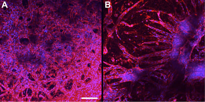
Figure 2. Microvessel networks fabricated with ultrasound standing wave fields. A) Capillary-like network formed with 0.1 MPa, 1 MHz ultrasound field. B) Branching arteriole/venule-like network formed with 0.3 MPa, 1 MHz ultrasound field. Images were collected using multiphoton immunofluorescence microscopy. Scale bar, 50 microns. (Garvin et al. 2013).
The use of ultrasound for microvascular tissue engineering is ideal as it is non-invasive, rapid, inexpensive, can be adapted to various dimensions, and does not affect cell viability. Furthermore, ultrasound fabrication processes can be broadly applied to many cell types and gel compositions and can be easily incorporated into commercial fabrication processes.
References:
[1] Garvin KA, Hocking DC, Dalecki D. Controlling the spatial organization of cells and extracellular matrix proteins in engineered tissues using ultrasound standing wave fields. Ultrasound Med. Biol. 36: 19191932; 2010.
[2] Garvin KA, Dalecki D, Hocking DC. Vascularization of three-dimensional collagen hydrogels using ultrasound standing wave fields. Ultrasound Med. Biol. 37: 1853-1864; 2011.
[3] Garvin KA, Dalecki D, Youssefhussien M. Helguera M, Hocking DC. Spatial patterning of endothelial cells and vascular network formation using ultrasound standing wave fields. J. Acoust. Soc. Am. 134: 1483-1490; 2013.