Fourth ASA/ASJ Joint Meeting, Honoloulu, Hawaii
Biomedical Application of Acoustic Microscopy
Yoshifumi Saijo - saijo@idac.tohoku.ac.jp
Popular version of paper 3pBB3
Brief History of Acoustic MicroscopyOptics has been the main observational mode in the microscopic world for many years. However, its domain is restricted to largely transparent media. Sokolov in the former Soviet Union, first proposed the concept of an ultrasonic microscope in order to visualize opaque media in 1949. He pointed out that the wavelength of 3-GHz ultrasound in water is the same as the wavelength of green light in air. At that time, however, the technologies to produce 3-GHz ultrasound did not exist. In 1959, Dunn and Fry at University of Illinois developed an ultrasonic absorption microscope method with 12-MHz ultrasound. During the 1970s, high frequency techniques were developed for the microwave technology for radar and for satellite communications. In 1973, Quate and Lemons at Stanford University developed a scanning acoustic microscope (SAM), now used in the biomedical field because of its high resolution and high quality imaging. Principles and Objectives of Classical Acoustic MicroscopyThe acoustic focusing element comprises a ZnO piezoelectric transducer with a sapphire lens. The ultrasonic frequency is variable over the range of 100 to 210 MHz and the beam width at the focal volume ranges from 5 mm (at 210 MHz) to 10 mm (at 100 MHz). The attenuation and sound speed of the material can be derived from the intensity and phase of the reflected signal, respectively. The attenuation depends on the molecular weight of the material. Sound speed has close correlation to the density and elastic bulk modulus. Thus, acoustic properties can be applied to assess the mechanical or physical properties of biological materials.
We have been developing SAM for medicine and biology since 1985. The objectives of the SAM study in medicine and biology are following. First, SAM can be applied for intra-operative pathological examinations since it does not require special staining techniques. Second, ultrasonic data obtained with the high frequency SAM can be used for assessing reflectability or texture in clinical echographic imaging. Third, the biomechanics tissues are measured by acoustic parameters because the sound speed has close correlation to the elastic bulk modulus.
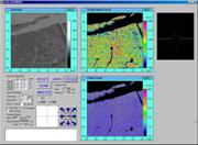
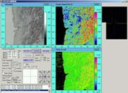
Each pixel in the acoustic microscope image shows the quantitative value of the acoustic parameters. The values are shown by the color bar scale. The arrows in the following images indicate 500 mm. Examples of Classical Acoustical Microscopy Images1) Myocardial Infarction [1]
2) Atherosclerosis [2-4]
3) Brain
4) Cells [5]
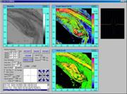
Sound Speed Microscopy [6,7]In 2002, we proposed a new concept acoustic microscope using a single pulsed ultrasound instead of burst waves in the conventional acoustic microscopy. The pulse response is analyzed in the frequency domain and the thickness, attenuation and speed of sound are measured at all the sampling points. Thanks for the development of computer and digital technology, the size of the instruments can be reduced to set on a desktop. Examples of Sound Speed Microscopy Images1) Atherosclerosis
2) Cardiomyopathy
3) Breast Cancer
Acoustic Impedance MicroscopyAs the specimens for classical acoustic microscopy and sound speed microscopy need to be flat and thin, the application was limited to the excised sample or cultured cells. In 2005, we proposed another new concept acoustic microscopy to observe the surface of the specimen without slicing. The system enables the in vivo high resolution imaging and can be applied for intra-operative examination
References
|