
Sounds of Cuvier's Beaked Whale and the Bottlenose Dolphin
Ted W. Cranford - tcranfor@mail.sdsu.edu
Biology Department, San Diego State University
Petr Krysl - pkrysl@ucsd.edu
Structural Engineering, University of California at San Diego,
John A. Hildebrand - jhildebrand@ucsd.edu
Marine Physical Lab, University of California San Diego,
Popular version of paper 4aAB5 and 4aAB4
Presented Thursday morning, May 21, 2009
157th ASA Meeting, Portland, OR
Investigating the physiology of sound reception in the largest animals in the world is problematic. Their gargantuan size and marine habitat often makes them difficult to find or access, and impossible to handle alive; quite simply, they are intractable subjects for study. The rapid advance of blazing fast computer hardware, giant industrial X-ray CT scanners, and modern mathematical and engineering approaches to building computer models has allowed us to pierce the seeming impenetrable veil around the bioacoustics of large whales.
The potential for sound to impact living marine resources has become an important topic in the last decade, initiated by stranded whales associated with Navy sonar, heightened public concern, and at least one court case rising all the way to the United States Supreme Court. Recently, evidence has surfaced that suggests human-generated sound can have detrimental effects on fish hearing, reproductive habits, and stress levels. We have developed a suite of techniques that combine to produce a “finite element model” of a whale’s head. This model allows us to simulate what happens when the anatomy of the whale interacts with sound. We are now capable of deciphering the physics and physiology of sound production and sound reception in the head of an adult male Cuvier’s beaked whale (Ziphius cavirostris).
The first step in the process was to develop a method for collecting a representation of in situ anatomic structure, the anatomic geometry, from large whales. The central key to the solution is rocket science! More specifically, the technology used to inspect solid fuel rockets for flaws, an industrial x-ray CT scanner, can be used to scan a dead whale. We waited for an opportunity when we knew a whale had recently beached itself alive and then subsequently died. By getting to the recently deceased behemoth and covering it with ice before it got too far along in the decomposition process, then we could salvage the specimen in good enough condition to scan it and build a computer model from its carcass.
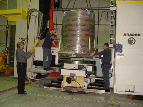 |
| Figure 1. Industrial x-ray CT scanner at Hill Air Force Base is normally used to scan solid fuel rocket motors. In this instance, it will scan a frozen whale head contained within a sarcophagus built specifically for this purpose. |
After scanning the head of a Cuvier’s Beaked Whale, we published a report about the structure of its biosonar anatomy. (Cranford TW, et al., (2008), Anatomic Geometry of Sound Transmission and Reception in Cuvier's Beaked Whale (Ziphius cavirostris). Anat Rec 291:353-378). That paper was the first quantitative description of biosonar anatomy for any toothed whale.
After the scanning process, we dissected the specimen, took it apart systematically, and measured the elastic properties of the various tissues. These values for tissue elasticity and the tissue density values given by the CT scans were the two primary building blocks for the computer model. The toolbox that took the building blocks and constructed the model is our finite element modeling software, the Vibroacoustic Toolkit, which was custom-built for this purpose. Computer simulations led to another published paper that reported the discovery a new channel for sound reaching the ears in Cuvier’s Beaked Whale. (Cranford TW, Krysl P, Hildebrand JA. (2008). Sound pathways revealed: Simulated sound transmission and reception in Cuvier’s beaked whale (Ziphius cavirostris). Bioinsp. Biomim. 3:1, 1-10.).
The computer simulations show that most sounds arriving from in front of the animal enter the head underneath the tongue region, pass through the throat and an opening in the posterior part of hollow lower jaw, and propagate along a fat body to the ear.
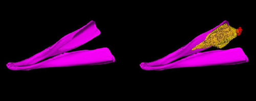 |
| Figure 2. These views of the jaws (magenta) from a Cuvier’s Beaked Whale are reconstructed from X-ray CT scans. The image on the left shows that the rear portion of the jaws is hollow and there is no bony wall on the inside. This is even more evident in the image on the right, in which the fat body (yellow) is clearly seen because the bony wall is missing. The ear complex (red) is also shown. |
In order for this sound reception pathway to function, the bony wall on the inside of the lower jaws must be absent; we say, “The door must be open.” As it turns out, all living toothed whales have this open door. And some of the earliest fossils also show the same hollow jaw with the open door. This suggests that this sound reception pathway developed early in the evolution of whales.
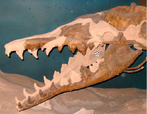 |
| Figure 3. Ancient whales are known as archaeocetes. The fossil whale shown in the above image is Basilosaurus cetoides. It lived approximately 35-40 million years ago. Notice that it has the same hollow (asterisk) in the rear portion of the lower jaw and, perhaps more importantly, the same missing wall of bone. |
The computer model allows us to visualize the pathway or “river” of sound as it “flows” from the front of the head to the ear complex. We call this the gular pathway. It passes through the throat region of the animal.
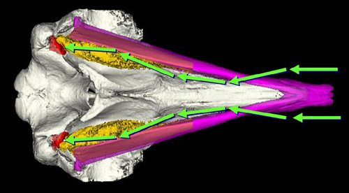 |
| Figure 4. This image shows a view from the underside of the head. The arrows show the generalized pathway (rivers) for sound passing from in front of Cuvier’s Beaked Whale, underneath the jaws (magenta), through the fat body (yellow), and to the ears (red). |
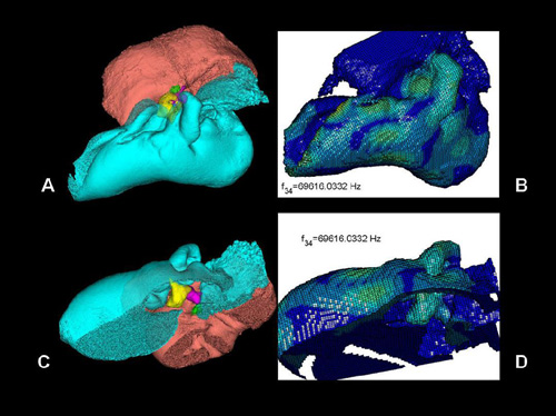 |
| Figure 5. These images show the anatomy of the bony ear complex (tympanoperiotic complex or TPC) and predictions for how it vibrates. Panel A shows a left lateral view of the TPC, the periotic bone (salmon), the tympanic bone (cyan), and the middle ear bones or ossicles (malleus = yellow, incus = magenta, stapes = green). Panel C shows the TPC upside down with one side removed to facilitate visualizing the ossicles (same color map). Panels B & D show the TPC subdivided into finite elements whose properties are known. The analysis predicts the vibrational patterns for the entire TPC at 69 kHz. The lighter colors indicate higher amplitude motions of elements. |
The computer model allows us to predict how the bony ear complex will vibrate as a result of being stimulated by the incoming river of sound. This is known as vibrational analysis. It describes how all the stiffness and mass features interact to produce the collective motion of the entire structure based on elementary physical principles.
The TPC collects sound from the gular pathway (river) and transmits it to the cochlea (inner ear). Analysis suggests that Navy sonar sounds reach the ears with reduced amplitude, but the frequencies used by these animals to catch prey are amplified.
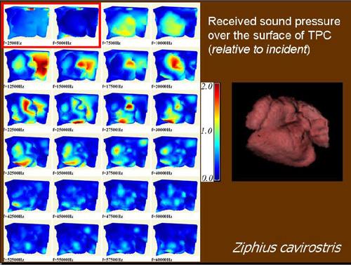 |
| Figure 6. This composite image tells us about the received sound pressure over the surface of the TPC with respect to the sound incident on the head. The image on the right shows the TPC of Cuvier’s Beaked Whale reconstructed from CT scans. The twenty four subpanels on the left show where the relative pressure “pushes” on the TPC after flowing through the river. Relative pressures are color coded (Green = 1 = pressure is equal to the sound pressure incident on the head. Blue = 0 = sound pressure is below incident pressure. Red = 2 = pressure is twice the incident pressure). Interestingly, the first two subpanels (outlined with a red line) indicate that the frequencies in the range produced by U.S. Navy’s Mid Frequency Active Sonar are largely filtered out (i.e. blue indicates less sound pressure than incident) before they reach the ear complex! The other subpanels (7.5 kHz to 42.5 kHz) show that those frequencies and locations that are particularly effective at driving the vibrations of the bony ear complex (TPC). These frequencies are also those that are produced by these animals in the wild for the purposes of biosonar. |
Computer models are powerful tools for investigation and discovery of bioacoustic physiology, providing a means to simulate across a broad range of scales and taxonomies, from whales to fish. Virtual experiments can also determine potential for and mechanism(s) of physical injury. Our techniques also provide potential for evaluating and directing mitigation efforts.
We acknowledge the continued support of Dr. Frank Stone and Dr. Ernie Young at the Chief of Naval Operations (N45); Dr. Curt Collins from the Naval Postgraduate school, as well as support from Dr. Mike Weise and Dr. Bob Gisiner at the Office of Naval Research.