
Xiaoming Zhang, PhD –Zhang.Xiaoming@mayo.edu
Department of Radiology
Mayo Clinic
Rochester, MN 55905
Alex X. Zhang, Xiaolei Xu, PhD
Department of Biochemistry and Molecular Biology
Mayo Clinic
Rochester, MN 55905
Popular version of paper 1aBAd2
Presented Monday morning, December 7, 2020
179th ASA Meeting, Acoustics Virtually Everywhere
Zebrafish are increasingly being used as animal models for human diseases such as cardiomyopathy and neuroblastoma. Like humans, zebrafish have a near-fully sequenced genome. However, the body of a zebrafish is only about 1.5-2.5 cm in length, which is much smaller than a person. To extrapolate results from zebrafish to humans, reliable quantitative measures on zebrafish are needed.
In this pilot study, we develop two noninvasive measurement techniques in zebrafish. One is to measure the heart function of zebrafish using echocardiography. Another is to measure the elastic property of zebrafish tissues using ultrasound vibro-elastography.
In zebrafish echocardiography, an adult zebrafish was anesthetized for three minutes in a tricaine solution. The zebrafish was then taken out of the anesthetic solution and positioned in a specially designed holder. The high-frequency Vevo 3100 ultrasound system with a MX700 ultrasound probe (29-71 MHz) was used to measure the heart function of the zebrafish. Figure 1 shows the experimental setup. Ultrasound imaging was used to measure heart volumes at the end of systole and diastole. The ejection fraction of the heart was analyzed. Pulse-wave Doppler was also used to analyze the heart function. We developed a technique to improve zebrafish echocardiography by removing the surface skin tissue near the heart of a zebrafish, which significantly improved the resolution of ultrasound images for analyzing heart function in zebrafish. All zebrafish recovered from this procedure and the subsequent echocardiography exam.
Another pilot study was to measure the elastic properties of zebrafish using ultrasound vibro-elastography. A 0.1 second gentle harmonic vibration was generated on the tail of a zebrafish using a sphere tip indenter with a 3 mm diameter. Shear wave propagation in the zebrafish was measured using another ultrasound system with a high frequency 18 MHz ultrasound probe. High frame rate ultrasound images were obtained using this ultrasound system to measure the generated wave propagation (300-500 Hz) in the bodies of the zebrafish. Figure 2 shows the experimental setup. Video 1 shows the wave propagation in a zebrafish. A region of interested (ROI) was used to analyze the sheer wave speed map. The ROI covered the most central area of the zebrafish surrounding the heart. The wave speed was 3.13 ± 1.20 (m/s) in the ROI at 300 Hz. It was found that wave speed increased from 300 Hz to 500 Hz as it passed through the zebrafish. All zebrafish recovered from this experiment. We will improve this technique for measuring elastic properties of the heart of zebrafish. It is feasible to develop this technique for measuring the elastic properties of zebrafish for phenotyping various diseases.
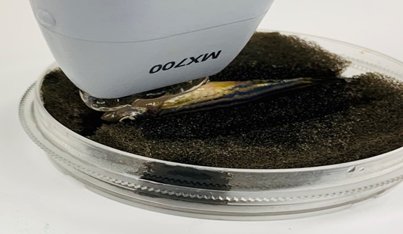 Figure 1. Experiment setup of zebrafish echocardiography.
 Figure 2. Experimental setup of zebrafish ultrasound vibro-elastography.