4pBAb3 – Bubbles on kidney stones in humans

Julianna C. Simon – jcsimon@psu.edu
Graduate Program in Acoustics
The Pennsylvania State University
201E Applied Science Building
University Park, PA 16802
AND
Center for Industrial and Medical Ultrasound
Applied Physics Laboratory
University of Washington
1013 NE 40th St.
Seattle, WA 98105

Popular version of paper 4pBAb3
Presented Thursday afternoon, December 5, 2019
178th ASA Meeting, San Diego, CA

Kidney stones affect 1 in 11 Americans, with an associated annual cost exceeding $10 billion. Most people are diagnosed with kidney stones using x-ray or CT when they go to the emergency room with severe side pain. Ultrasound can also be used, but the greyscale images can be difficult to interpret. Even though Doppler ultrasound is usually used to monitor blood flow, when you image a kidney stone with Doppler ultrasound, it appears as rapidly changing color, called the twinkling artifact (Fig. 1). This can enhance kidney stone detection with ultrasound, but it doesn’t appear on all stones. Recently, bubbles on the stone were suggested to cause the twinkling artifact because small changes in pressure influence the appearance of twinkling. However, these studies were done on stones outside of the human body, where exposure to air could artificially introduce the bubbles.

kidney stones

Fig. 1: The color Doppler ultrasound twinkling artifact highlights a kidney stone with rapidly-changing color.

In this presentation, we look at the origin of the twinkling artifact on kidney stones by imaging kidney stones in humans with ultrasound while inside a pressure or hyperbaric chamber (Fig. 2). Seven human subjects were exposed to 4 atmospheres of pressure while imaging with ultrasound. We found that twinkling was reduced by 39% at 4 atmospheres compared to twinkling at atmospheric pressure, which was statistically significant. This result suggests, for the first time, that bubbles exist in humans on the surface of kidney stones!

kidney stones

Fig. 2: Imaging people with kidney stones inside a hyperbaric chamber. Due to the risk of fire, the ultrasound system was outside the chamber with the transducer inserted through a port.

We were also curious as to whether these bubbles existed only on the stone surface, or whether they could be embedded in the stone during the stone formation process. In the lab, stones that had been removed from humans were imaged with high resolution CT while reducing the pressure around the stone. We found regions within the stone that grew when we reduced the pressure (Fig. 3), suggesting that bubbles can exist inside the stone, too.

Fig. 3: A high-resolution CT scan of a human kidney stone shows a small, dark region within a crack that expands when the ambient pressure is reduced (yellow circle), suggesting the bubbles are contained within the kidney stone.

NASA funded the study because they are interested in ultrasound for spaceflight and have found that changes in gas composition on space vehicles and pressure in spacesuits affects bubbles associated with decompression sickness and ultrasound imaging. Spaceflight increases the risk of forming kidney stones because bones demineralize, releasing calcium into the blood that is filtered through the kidney. Only four times have astronauts prepared for emergency return to Earth, and one was for a stone that eventually passed. Because of the risk to astronauts and people on Earth, both NASA and NIH have funded researchers at the University of Washington to develop an ultrasound system to image, fragment, and expel stones from the kidney. The key to using this system is being able to see the stone with ultrasound, which is where this work on understanding the twinkling artifact plays an important role.

3pBA4 – Artificial Intelligence for Automatic Tracking of the Tongue in Real-time Ultrasound Data

M. Hamed Mozaffari – mmoza102@uottawa.ca
Won-Sook Lee – wslee@uottawa.ca
School of Electrical Engineering and Computer Science (EECS)
University of Ottawa
800 King Edward Avenue
Ottawa, Ontario, Canada K1N 6N5

David Sankoff – sankoff@uottwa.ca
Department of Mathematics and Statistics
University of Ottawa
150 Louis Pasteur Pvt.
Ottawa, Ontario K1N 6N5

Popular version of papers 3pBA4
Presented Wednesday afternoon, December 4, 2019
178th ASA Meeting, San Diego, CA

Medical ultrasound technology has been a well-known method in speech research for studying of tongue motion and speech articulation. The popularity of ultrasound imaging for tongue visualization is because of its attractive characteristics such as imaging at a reasonably rapid frame rate, which allows researchers to visualize subtle and swift gestures of the tongue during the speech in real-time. Moreover, ultrasound technology is relatively affordable, portable and clinically safe with a non-invasive nature.

Exploiting the dynamic nature of speech data from ultrasound tongue image sequences might provide valuable information for linguistics researchers, and it is of great interest in many recent studies. Ultrasound imaging has been utilized for tongue motion analysis in the treatment of speech sound disorders, comparing healthy and impaired speech production, second language training and rehabilitation, to name a few.

During speech data acquisition, an ultrasound probe under the user’s jaw pictures tongue surface in midsagittal or coronal view in real-time. Tongue dorsum can be seen in this view as a thick, long, bright, and continues region due to the tissue-air reflection of ultrasound signal by the air around the tongue. Due to the noise characteristic of ultrasound images with the low-contrast property, it is not an easy task for non-expert users to localize the tongue surface.

tongue

Picture 1: An illustration of the human head and tongue mid-sagittal cross-section view. The tongue surface in ultrasound data can be specified using a guide curve. Highlighted lines (red and yellow) can help users to track the tongue in real-time easier.

To address this difficulty, we proposed a novel artificial intelligence method (named BowNet) for tracking the tongue surface in real-time for non-expert users. Using BowNet, users can see a highlighted version of their tongue surface in real-time during a speech without any training. This idea of tracking tongue using a contour facilitates linguistics to use the BowNet technique for their quantitative studies.

Performance of BowNet in terms of accuracy and automation is significant in comparison with similar methods as well as the capability of applying on different ultrasound data types. The real-time performance of the BowNet enables researchers to propose new second language training methods. The better performance of BowNet techniques is presented in Video 1.

Video1: A performance presentation of BowNet models in comparison to similar recent ideas. Better generalization over different datasets, less noise, and better tongue tracking can be seen. Failure cases with colour are indicated in video.

2pBA2 – Double, Double, Toil and Trouble: Nitric Oxide or Xenon Bubble

Christy K. Holland – Christy.Holland@uc.edu
Department of Internal Medicine, Division of Cardiovascular Health and Disease and
Department of Biomedical Engineering
University of Cincinnati
Cardiovascular Center 3935
231 Albert Sabin Way
Cincinnati, Ohio  45267-0586
https://www.med.uc.edu/ultrasound
office:  +1 513 558 5675

Himanshu Shekhar – h.shekhar.uc@gmail.com
Department of Electrical Engineering
AB 6/327A
Indian Institute of Technology (IIT) Gandhinagar
Palaj 382355, Gujarat, India

Maxime Lafond – lafondme@ucmail.uc.edu
Department of Internal Medicine, Division of Cardiovascular Health and Disease and
Department of Biomedical Engineering
University of Cincinnati
Cardiovascular Center 3933
231 Albert Sabin Way
Cincinnati, Ohio  45267-0586

Popular version of paper 2pBA2
Presented Tuesday afternoon at 1:20 pm, May 14, 2019
177th ASA Meeting, Louisville, KY

Designer bubbles loaded with special gases are under development at the University of Cincinnati Image-guided Ultrasound Therapeutics Laboratories to treat heart disease and stroke. Xenon is a rare, pricey, heavy, noble gas, and a potent protector of a brain deprived of oxygen. Nitric oxide is a toxic gas that paradoxically plays an important role in the body, triggering the dilation of blood vessels, regulating the release and binding of oxygen in red blood cells, and even killing virus-infected cells and bacteria.

Microbubbles loaded with xenon or nitric oxide stabilized against dissolution with a fatty coating, can be exposed to ultrasound for site-specific release of these beneficial gases, as shown in the video (Supplementary Video 1). The microbubbles were stable against dissolution for for 30 minutes, which is longer than the circulation time before removal from the body. Curiously, the co-encapsulation of either of these bioactive gases with a heavier perfluorocarbon gas increased the stability of the microbubbles. Bioactive gas-loaded microbubbles act as a highlighting agent on a standard diagnostic ultrasound image (Supplementary Video 2). Triggered release was demonstrated with pulsed ultrasound already in use clinically. The total dose of xenon or nitric oxide was measured after release from the microbubbles. These results constitute the first step toward the development of ultrasound-triggered release of therapeutic gases to help rescue brain tissue during stroke.

Supplementary Video 1: High-speed video of a gas-loaded microbubble exposed to a single Doppler ultrasound pulse. Note the reduction in size over exposure to ultrasound, thus demonstrating acoustically-driven diffusion of gas out of the microbubble.

Supplementary Video 2: Ultrasound image of a rat heart filled with nitric oxide-loaded microbubbles. The chamber of the heart appears bright because of the presence of the microbubbles.

4aBAa7 – Unprecedented high-spatial resolution was achieved in ultrasound imaging by breaking the fundamental limitation with the operating ultrasound wavelength

Kang Kim – kangkim@upmc.edu
Qiyang Chen – qic41@pitt.edu
Jaesok Yu – jaesok.yu@ece.gatech.edu
Roderick J Tan – tanrj@upmc.edu
University of Pittsburgh
3550 Terrace St, room 623, Pittsburgh, PA 15261

Popular version of paper 2aBA8; 4aBAa7
Presented Tuesday & Thursday morning, May 14 & 16, 2019
177th ASA Meeting, Louisville, KY

US imaging is one of the most favored imaging modalities in clinics in general because of its real-time display, safety, noninvasiveness, portability and affordability. One major disadvantage of ultrasound imaging is its limited spatial resolution that is fundamentally governed by the wavelength of the operating ultrasound. We developed a new super-resolution imaging algorithm that can achieve super high-spatial resolution beyond such limitation called acoustic diffraction limit.

The concept of the super resolution that bypasses a physical limit for the maximum resolution of traditional optical imaging was originally introduced in microscopy imaging community and later developed into a ground-breaking technology of the nano-dimension microscopy imaging, for which the Nobel Prize in Chemistry was awarded in 2014. In brief, microscopy super resolution imaging technology is based on randomly repeated blinking process of the fluorophores in response to the light source of the microscopy. In recent years, the concept has been translated into ultrasound imaging community. The random blinking process that requires for achieving super resolution using ultrasound is provided by flowing microbubbles in blood vessels which randomly oscillate in response to the ultrasound pressure from the imaging transducer. The maximum spatial resolution in super resolution microscopy technology is in the range of tens of nanometers (10-9 m) that allows to visualize the pathways of individual molecules inside living cells, while ultrasound super resolution imaging can achieve a spatial resolution in the range of tens of micrometers (10-6 m) when using a typical clinical ultrasound imaging transducer of a few MHz center frequency. However, due to the large imaging depth of ultrasound up to several centimeters, ultrasound super resolution imaging technology is practically very useful in imaging human subject with greater details of microvasculature which is of critical importance for many diseases.

Figure 1

Traditional contrast enhanced ultrasound (CEU) imaging technologies using microbubbles provide superior contrast of vasculatures, effectively suppressing the surrounding tissue signals, but the spatial resolution remains to the acoustic diffraction limit. In recent years, to overcome such limitation with CEU, several approaches have been made to overcome such limitation by employing super resolution concept, however requiring a long scan time, which hinders the technology from being wide spread. The major contribution from my laboratory is to drastically shorten the scan time of super resolution imaging using deconvolution algorithm for microbubble center localization, as well as to compensate artifacts due to physiological motions using block matching based motion correction and spatio-temporal-interframe-correlation based data re-alignment, so that the technology can be used in vivo for diverse applications. In brief, a novel approach of ultrafast ultrasound imaging, rigid motion compensation, tissue signal suppressor and deconvolution based deblurring has been developed for both high spatial and temporal resolution.

Video 1

The developed technology was applied in imaging microvasculature change which is a critical feature during disease development and progress. Vasa vasorum that is network of small blood vessels that supply the walls of large blood vessels and often multiplies and infiltrates into atherosclerotic plaque were identified in rabbit model.

Figure2

Microvascular rarefaction is a key signature of acute kidney injury that often progress into chronic kidney diseases and eventual kidney failure. Microvessels in mouse acute kidney injury model were successfully identified and quantitatively analyzed.

Figure 3

1pBA11 – An ultrasound surface wave elastography technique for noninvasive measurement of scar tissue

Boran Zhou – zhou.boran@mayo.edu
Xiaoming Zhang – zhang.xiaoming@mayo.edu
Department of Radiology, Mayo Clinic,
Rochester, MN 55905

Saranya P. Wyles – Wyles.Saranya@mayo.edu
Alexander Meves – Meves.Alexander@mayo.edu
Department of Dermatology, Mayo Clinic,
Rochester, MN 55905

Steven Moran – Moran.Steven@mayo.edu
Department of Plastic Surgery, Mayo Clinic,
Rochester, MN 55905

Popular version of paper 1pBA11
Presented Monday afternoon, May 13, 2019
177th ASA Meeting, Louisville, KY

Hypertrophic scars and keloids are characterized by excessive fibrosis and can be functionally problematic. Indeed, hypertrophic scarring is characterized by wide, raised scars that remain within the original borders of injury and have a rapid growth phase. We have developed an ultrasound elastography technique to assess the skin elasticity for patients with scleroderma (1). Currently, no clinical technique is available to noninvasively quantify and assess the progression and development of scar restoration. There is a need for quantitative scar measurement modalities to effectively evaluate and monitor treatments.

We aim to assess the role of ultrasound surface wave elastography (USWE) in accurately evaluating scar metrics. 3 Patients were enrolled in this research based on their clinical diagnoses. For the patients with scar on the forearm, they were tested in a sitting position with their left or right forearm or upper arm placed horizontally on a pillow in a relaxed state. The indenter of the handheld shaker was placed on the tissue at control and scar sites. A 0.1-s harmonic vibration was generated by the indenter on the tissue (2). The vibration was generated at 3 frequencies: 100, 150 and 200 Hz. An ultrasound system with an ultrasound probe with a central frequency of 6.4 MHz was positioned about 5 mm away from the indenter and used for detecting the surface wave motion of the tissue (3).

The wave motions on the 8 selected locations on the tissue surface were noninvasively measured using our ultrasound-based method (Fig. 1a)(4). The phase change with distance of the harmonic wave propagation on the tissue surface was used to measure the surface wave speed.
The measurement of wave speed can be improved by using multiple phase change measurements over distances (5). The regression of the phase change with distance can be obtained by “best fitting” a linear relationship between them (Fig. 1b). Using the tissue motion at the first location as a reference, the wave phase delay of the tissue motions at the remaining locations, relative to the first location, was used to measure surface wave speed (6).

Wave speeds of forearm or upper arm control and scar sites of the 3 patients at 100, 150, and 200 Hz before and after treatment were compared in Figure 2. The p values for the t-tests of the differences between before and after treatment were less than 0.05 for scar sites at 3 frequencies. The higher wave speed indicates the stiffer tissue. The obtained results suggest that scar portion was softener after treatment. USWE provides an objective assessment of the reaction of the scar to injury and treatment response.

Figure 1. (a) Representative B-mode image of skin, (b) Blue circles represent the selected dots for wave speed measurement.

Figure 2. Comparison of wave speeds at 3 frequencies between forearm control and scar sites.
References

1. Zhang X, Zhou B, Kalra S, Bartholmai B, Greenleaf J, Osborn T. An Ultrasound Surface Wave Technique for Assessing Skin and Lung Diseases. Ultrasound in Medicine & Biology. 2018;44(2):321-31.
2. Zhang X, Osborn T, Zhou B, et al. Lung ultrasound surface wave elastography: a pilot clinical study. IEEE transactions on ultrasonics, ferroelectrics, and frequency control. 2017;64(9):1298-304.
3. Clay R, Bartholmai BJ, Zhou B, et al. Assessment of Interstitial Lung Disease Using Lung Ultrasound Surface Wave Elastography: A Novel Technique With Clinicoradiologic Correlates. J Thorac Imaging. 2018.
4. Zhang X, Zhou B, Miranda AF, Trost LW. A Novel Noninvasive Ultrasound Vibro-elastography Technique for Assessing Patients With Erectile Dysfunction and Peyronie Disease. Urology. 2018;116:99-105.
5. Zhou B, Zhang X. The effect of pleural fluid layers on lung surface wave speed measurement: Experimental and numerical studies on a sponge lung phantom. Journal of the Mechanical Behavior of Biomedical Materials. 2019;89:13-8.
6. Zhang X, Zhou B, Osborn T, Bartholmai B, Kalra S. Lung ultrasound surface wave elastography for assessing interstitial lung disease. IEEE Transactions on Biomedical Engineering. 2018:1-.

4aSP4 – Streaming Video through Biological Tissues using Ultrasonic Communication

Gizem Tabak – tabak2@illinois.edu
Michael Oelze – oelze@illinois.edu
Andrew Singer – acsinger@illinois.edu
University of Illinois at Urbana-Champaign
306 N Wright St
Urbana, IL 61801

Popular version of paper 4aSP4
Presented Thursday morning, May 16, 2019
177th ASA Meeting, Louisville, KY

Researchers at the University of Illinois at Urbana-Champaign have developed a fast, wireless communication alternative that also has biomedical implications. Instead of using radio frequency (RF) to transmit signals, the team is using ultrasonic waves to send signals at high enough data rates to transmit video through animal or human tissue.

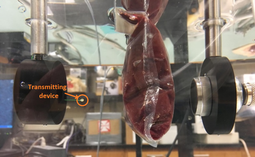
The team of electrical and computer engineering professors Andrew Singer and Michael Oelze and graduate researcher Gizem Tabak have achieved a transmission rate of 4 megabits per second through animal tissue with 2-mm transmitting devices. This rate is high enough to send high definition video (3 Mbps) and 15 times faster than that RF waves can currently deliver.

ultrasonic communication

Figure 1 – Experimental setup for streaming at 4Mbps through 2” beef liver

The team is using this approach for communicating with implanted medical devices, like those used to scan tissue in a patients’ gastrointestinal (GI) tract.

Currently one of two methods are used to image the GI tract. The first is video endoscopy, which involves inserting a long probe with a camera and light down the throat to take real-time video and send it to an attached computer. This method has limitations in that it cannot reach the midsection of the GI tract and is highly invasive.

The second method involves a patient swallowing a pill that contains a mini camera that can take images throughout the tract. After a day or so, the pill is retrieved, and the physician can extract the images. This method, however, is entirely offline, meaning there is no real-time interaction with the camera inside the patient.

A third option uses the camera pill approach but sends the images through RF waves, which are absorbed by the surrounding tissue. Due to safety regulations governing electromagnetic radiation, the transmitted signal power is limited, resulting in data rates of only 267 kilobits per second.

The Illinois team is proposing to use ultrasound, a method that has already proven safe for medical imaging, as a communication method. Having achieved data rates of 4 Mbps with this system through animal tissue, the team is translating the approach to operate in real-time for use in the human body.

Pairing this communication technology with the camera pill approach, the device not only could send real-time video, but also could be remotely controlled. For example, it might travel to specific areas and rotate to arbitrary orientations. It may even be possible to take tissue samples for biopsy, essentially replacing endoscopic procedures or surgeries through such mini-remote controlled robotic devices.