Needle-Free Ultrasound Vaccine Delivery #Acoustics23

Needle-Free Ultrasound Vaccine Delivery #Acoustics23

Technique employs bubbles formed and popped in response to sound waves to deliver vaccines and achieve potentially improved immune response.

SYDNEY, Dec. 4, 2023 – An estimated quarter of adults and two-thirds of children have strong fears around needles, according to the U.S. Centers for Disease Control and Prevention. Yet, public health depends on people being willing to receive vaccines, which are often administered by a jab.

Darcy Dunn-Lawless, a doctoral student at the University of Oxford’s Institute of Biomedical Engineering, is investigating the potential of a painless, needle-free vaccine delivery by ultrasound. He will share the recent advancements in this promising technique as part of Acoustics 2023 Sydney, running Dec. 4-8 at the International Convention Centre Sydney. His presentation will take place Dec. 4 at 11:00 a.m. Australian Eastern Daylight Time.

“Our method relies on an acoustic effect called ‘cavitation,’ which is the formation and popping of bubbles in response to a sound wave,” said Dunn-Lawless. “We aim to harness the concentrated bursts of mechanical energy produced by these bubble collapses in three main ways. First, to clear passages through the outer layer of dead skin cells and allow vaccine molecules to pass through. Second, to act as a pump that drives the drug molecules into these passages. Lastly, to open up the membranes surrounding the cells themselves, since some types of vaccine must get inside a cell to function.”

Though initial in vivo tests reported 700 times fewer vaccine molecules were delivered by the cavitation approach compared to conventional injection, the cavitation approach produced a higher immune response. The researchers theorize this could be due to the immune-rich skin the ultrasonic delivery targets in contrast to the muscles that receive the jab. The result is a more efficient vaccine that could help reduce costs and increase efficacy with little risk of side effects.

“In my opinion, the main potential side effect is universal to all physical techniques in medicine: If you apply too much energy to the body, you can damage tissue,” Dunn-Lawless said. “Exposure to excessive cavitation can cause mechanical damage to cells and structures. However, there is good evidence that such damage can be avoided by limiting exposure, so a key part of my research is to try and fully identify where this safety threshold lies for vaccine delivery.”

Vaccine

Ultrasound pulses deliver vaccines through the skin without needles. This technique, which employs sound waves to create bubbles that forge a path for the vaccine, may be especially helpful for DNA vaccines. Credit: Darcy Dunn-Lawless

Dunn-Lawless works as part of a larger team under the supervision of Dr. Mike Gray, Professor Bob Carlisle, and Professor Constantin Coussios within Oxford’s Biomedical Ultrasonics, Biotherapy and Biopharmaceuticals Laboratory (BUBBL). Their cavitation approach may be particularly conducing to DNA vaccines that are currently difficult to deliver. With cavitation able to help crack open the membranes blocking therapeutic access to the cell nucleus, the other advantages of DNA vaccines, like a focused immune response, low infection risk, and shelf stability, can be better utilized.

###

Contact:
AIP Media
301-209-3090
media@aip.org

———————– MORE MEETING INFORMATION ———————–

The Acoustical Society of America is joining the Australian Acoustical Society to co-host Acoustics 2023 Sydney. This collaborative event will incorporate the Western Pacific Acoustics Conference and the Pacific Rim Underwater Acoustics Conference.

Main meeting website: https://acoustics23sydney.org/
Technical program: https://eppro01.ativ.me/src/EventPilot/php/express/web/planner.php?id=ASAFALL23

ASA PRESS ROOM
In the coming weeks, ASA’s Press Room will be updated with newsworthy stories and the press conference schedule at https://acoustics.org/asa-press-room/.

LAY LANGUAGE PAPERS
ASA will also share dozens of lay language papers about topics covered at the conference. Lay language papers are summaries (300-500 words) of presentations written by scientists for a general audience. They will be accompanied by photos, audio, and video. Learn more at
https://acoustics.org/lay-language-papers/.

PRESS REGISTRATION
ASA will grant free registration to credentialed and professional freelance journalists. If you are a reporter and would like to attend the meeting or virtual press conferences, contact AIP Media Services at media@aip.org. For urgent requests, AIP staff can also help with setting up interviews and obtaining images, sound clips, or background information.

ABOUT THE ACOUSTICAL SOCIETY OF AMERICA
The Acoustical Society of America (ASA) is the premier international scientific society in acoustics devoted to the science and technology of sound. Its 7,000 members worldwide represent a broad spectrum of the study of acoustics. ASA publications include The Journal of the Acoustical Society of America (the world’s leading journal on acoustics), JASA Express Letters, Proceedings of Meetings on Acoustics, Acoustics Today magazine, books, and standards on acoustics. The society also holds two major scientific meetings each year. See https://acousticalsociety.org/.

ABOUT THE AUSTRALIAN ACOUSTICAL SOCIETY
The Australian Acoustical Society (AAS) is the peak technical society for individuals working in acoustics in Australia. The AAS aims to promote and advance the science and practice of acoustics in all its branches to the wider community and provide support to acousticians. Its diverse membership is made up from academia, consultancies, industry, equipment manufacturers and retailers, and all levels of Government. The Society supports research and provides regular forums for those who practice or study acoustics across a wide range of fields The principal activities of the Society are technical meetings held by each State Division, annual conferences which are held by the State Divisions and the ASNZ in rotation, and publication of the journal Acoustics Australia. https://www.acoustics.org.au/

Using ultrasound as an antibody in Alzheimer’s and as a drug dose enhancer in cancer patients

Elisa Konofagou – ek2191@columbia.edu

Columbia University, 1210 Amsterdam Ave, New York, New York, 10027-7003, United States

Popular version of 2aBAa1 – Neuronavigated focused ultrasound for clinical bbb opening in alzheimer’s and brain cancer patients
Presented at the 184 ASA Meeting
Read the abstract at https://doi.org/10.1121/10.0018295

Ultrasound is widely known as an imaging modality in obstetrics and cardiology as well as several other applications but less known regarding its therapeutic effects despite its recent approvals in the clinic for ablation of prostate cancer and essential tremors. In the studies presented, we demonstrate that focused ultrasound (FUS) can be used in conjunction with microbubbles to open the blood-brain barrier (BBB) through the intact scalp of Alzheimer’s and pediatric tumor patients. The BBB is the main defense of the brain against toxic molecules but also prevents drugs from treating brain disease. In the case of Alzheimer’s, we demonstrate for the first time that the BBB opening resulting from FUS in the prefrontal cortex acts as an antibody in the brain. BBB opening results into a beneficial immune response in the brain that significantly reduces the beta amyloid in the region where ultrasound opened the blood-brain barrier. This was shown in 5 patients with Alzheimer’s.

In the case of the pediatric tumor patients, we aimed into the stem, which is a critical region between the spinal cord and the brain. The tumors in the pediatric patients are gliomas that grow in the stem where critical nerve fibers run through and they are therefore inoperable. We showed for the first time that BBB opening can be repeatedly induced with FUS in conjunction with microbubbles safely and efficiently in patients with pediatric glioma tumors in the stem. In this case, we used FUS in conjunction with a drug that, when crossing the blood-brain barrier, increases its efficiency. The patients reported smoother limb movement after treatment with the drug potentially acting more potently on the tumor.

It was concluded that ultrasound can safely open the blood-brain barrier in both patients as young as 6 years old to as old as 83 years old completely noninvasively and more importantly reduce the disease pathology and/or symptoms. The system is thus versatile, does not require a dedicated MR system or to be performed in the MR scanner unlike other systems and the entire procedure can last less than 30 min from start to finish. Ultrasound can thus be used alone or in conjunction with a drug in order to change the current dire landscape of treatment of brain disease. Finally, we show how Alzheimer’s beta amyloid and tau are excreted from the brain and can be detected with a simple blood test.

Ultrasonics to monitor liquid metal melt pool dynamics for improving metal 3D printing

Christopher Kube – kube@psu.edu
Twitter: @_chriskube

Penn State University, 212 Earth and Engineering Sciences Bldg, University Park, PA, 16802, United States

Tao Sun, University of Virginia
Samuel Clark, Advanced Photon Source, Twitter: @advancedphoton

Find the authors on LinkedIn:
www.linkedin.com/in/chriskube
www.linkedin.com/in/suntao

Popular version of 3pID2-Acoustics for in-process melt pool monitoring during metal additive manufacturing, presented at the 183rd ASA Meeting.

3D printed or additively manufactured (AM) metal parts are disrupting the status quo in a variety of industries including defense, transportation, energy, and space exploration. Engineers now design and produce customizable parts unimaginable only a decade ago. New geometrical or part shape freedom inherent to AM has already led to part performance often beyond traditionally manufactured counterparts. In the years to come, another revolutionary performance jump is expected by enabling the AM process to control the grain layout and structural features on the microscopic scale. Grains are the building blocks of metal parts that dictate many of the performance metrics associated with the descriptors of bigger, faster, and stronger.

The second performance revolution of AM metal parts requires uncovering new knowledge in the complicated physics present during the AM process. 3D printed metals are born from an energy source such as a laser or electron beam to selectively melt feedstock material at microscopic locations dictated by the computerized part drawing. Melted locations temporarily form liquid metal melt pools that solidify after the energy source moves to another location. Resulting grain structure and pore/defect formation strongly depends on how the melt pool cools and solidifies.

Over the past five years, high-energy X-rays only available at particle accelerators are used for direct real-time visualization of AM melt pool dynamics and solidification. Figure 1 shows an example X-ray frame, which captured a laser-generated melt pool moving in a single direction with a speed of 800 mm/ms.


MATLAB Handle Graphics – click here to watch the video.

This situation mimics the laser and melt pool movement found during 3D printing metal parts. Being able to directly observe melt pool behavior has led to new and improved understanding of the underlying physics. Unfortunately, experiments at such X-ray sources is difficult to ascertain because of extremely high demand across the sciences. Additionally, the measurement technique relegated to high-energy X-ray sources is not transferrable to metal 3D printers that exist in normal industrial settings. For these reasons, ultrasonics are being explored as a melt pool monitoring technology that can be deployed within real 3D printers.

Ultrasound is commonly used for imaging and detecting features inside of solid materials. For example, ultrasound is applied in medical settings during pregnancy or for diagnostics. Application of ultrasound for melt pool monitoring is made possible because of the tendency of ultrasound to scatter from the melt pool’s solid/liquid boundary. The development of the technique is being supported alongside X-ray imaging at the Advanced Photon Source at Argonne National Laboratory. X-ray imaging is providing the extremely important ground truth melt pool behavior allowing for easy interpretation of the ultrasonic response. In Figure 1, the ultrasonic response from the exact same melt pool given in the X-ray video is being shown for two different sensors. As the melt pool enters the field of view of the ultrasonic sensors (see online video), features in the ultrasound response confirms their sensitivity to the melt pool.

In this research, high-energy X-rays are being used to develop the ultrasonic technique and technology. In the coming year, the knowledge developed will be leveraged such that ultrasound can be applied on its own for melt pool monitoring in real metal 3D printers. Currently, no existing technology can capture the highly dynamic melt pool behavior through the depth of the part or substrate.

Practical benefits and value of melt pool monitoring within 3D printers are significant. Ultrasound can provide a quick check to determine the optimal laser power and speed combinations toward accelerated determination of process parameters. Currently, determination of the optimal process parameters requires destructive postmortem microscopy techniques that are extremely costly, time-consuming (sometimes more than a year), and wasteful. Ultrasound has the potential to reduce these factors by an order of magnitude. Furthermore, metal 3D printing processes are highly variable over many months, across different machines, and even when using feedstock powder from different suppliers. Ultrasonic melt pool monitoring can provide period checks to assure variability is minimized.

2aPAb – Ultrasound technology to remove kidney stones

Mohamed A. Ghanem – mghanem@uw.edu
Adam D.  Maxwell – amax38@uw.edu
Oleg A. Sapozhnikov – olegs@uw.edu
Michael R. Bailey – mbailey@uw.edu

University of Washington
1013 NE 40th St.
Seattle WA 98105

Popular version of 2aPAb – Designing an array for acoustic manipulation of kidney stones
Presented Tuesday morning, May 24, 2022
182nd ASA Meeting
Click here to read the abstract

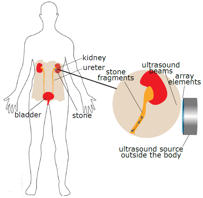
Ultrasound technology is becoming an important treatment tool. For instance, sound waves can apply a radiation pressure that can displace an object. Multi-element arrays are complex ultrasound sources that consist of several small transducers that can be driven in sync or a specific order to output pressure waves with different shapes. Pressure wave shapes that have a doughnut shape or a long tube are useful as they can trap an object in the center and as we control the location of the doughnut the object follows. This technology can be used to trap small kidney stones or stone fragments and move them from the kidney collection areas toward the kidney exit without surgery. We have demonstrated the ability to move kidney stone models in the bladders transcutaneously in live pigs under anesthesia. We are currently designing a new multi-element array that will enable us to adapt this technology to move stones in the complex structure of the kidney over larger distances. This technology will reduce the surgery associated with kidney stone treatments by removing small stones or fragments before they become larger, which will lead to surgery, and eliminating emergency room visits by relieving blockages from these stones or fragments.

kidney stones

Controlled steering of kidney stones toward  the kidney exit with an ultrasound array.

2aBAb2 – Feasibility of using ultrasound with microbubbles to purify cell lines for immunotherapy application

Thomas Matula – matula@uw.edu
Univ. of Washington
1013 NE 40th St.
Seattle, WA 98105

Oleg A. Sapozhnikov
Ctr. for Industrial and Medical Ultrasound
Appl. Phys. Lab
Univ. of Washington
Seattle, Washington
Phys. Faculty

Lev Ostrovsky
Dept. of Appl. Mathematics
University of Colorado
Inst. of Appl. Phys.
Russian Acad. of Sci.
Boulder, CO

Andrew Brayman
John Kucewicz
Brian MacConaghy
Dino De Raad
Univ. of Washington
Seattle, WA

Popular version of paper 2aBAb2
Presented Tuesday morning, Nov 6, 2018
176th ASA Meeting, Victoria, BC, Canada

Cells are isolated and sorted for a variety of diagnostic (e.g., blood tests) and therapeutic (e.g., stem cells, immunotherapy) applications, as well as for general research. The workhorses in most research and commercial labs are fluorescently-activated cell sorters (FACS) [1] and magnetically-labeled cell sorters (MACS) [2]. These tools use biochemical labeling to identify and/or sort cells which express specific surface markers (usually proteins). FACS uses fluorophores that target specific cell markers. The detection of a specific fluorescence wavelength tells the system to sort those cells. FACS is powerful and can sort based on several different cellular markers. However, FACS is also very expensive and complicated such that they are mostly found only in large core facilities.

MACS uses magnetic beads that attach to cell markers. Permanent magnets can then be used to separate magnetically-tagged cells from untagged cells. MACS is much less expensive than FACS, and can be found in most labs. However, MACS also suffers from weaknesses, such as low throughput, and can only sort based on a single marker.

We describe a new method that merges biochemical labeling with ultrasound-based separation. Instead of lasers and fluorophore tags (i.e., FACS), or magnets and magnetic particle tags (i.e., MACS), our technique uses ultrasound and microbubble tags (Fig. 1). Like FACS and MACS, we attach a biochemical label (an antibody) to attach a microbubble to the cell’s surface protein. We then employ an ultrasound pulse that generates an acoustic radiation force, pushing the microbubbles; the attached cells are dragged along with the microbubbles, effectively separating them from untagged cells. This is accomplished because cells only very lightly interact with ultrasound, whereas microbubbles interact very significantly with the sound waves. We theorized that the force acts on the microbubble while the cell acts as a fluid that adds a viscous drag to the system (see [3]).

immunotherapy

Figure 1. Cell separation technologies

We can break down our studies into two categories, cell rotation and cell sorting. In both cases we constructed an apparatus to view cells under a microscope. Figure 2 shows a cell rotating as the attached microbubbles align with the sound field (the movie can be found by clicking here). We developed a theory to describe this rotation, and the theory fits the data well, allowing us to ‘measure’ the acoustic radiation force on the conjugate microbubble-cell system (Fig. 3).

Figure 2. A leukemia cell has two attached microbubbles. An ultrasound pulse from above causes the cell to rotate.

Figure 3. We assume that the microbubbles act as point forces. The projection of these forces perpendicular to the radiation force direction leads to a torque on the cell, which is balanced by the viscous torque. This leads to an equation of motion that can be put in terms of angular displacement. Thus, the parameters are detailed in [3]. The results are plotted along with the data, showing a nice match between the theory and data. For our conditions, the acoustic radiation force was found to be F=1.7×10⁻¹²N. [IMAGE MISSING]

When placed in a flow stream with other cells, the tagged cells can be easily pushed with ultrasound. Figure 4a shows how a single leukemia cell is pushed downward while normal erythrocytes (red blood cells) continue flowing in the stream (the movie can be found by clicking here). This shows that one can effectively separate tagged cells. However, in a commercial setting, one wants to sort with a much higher concentration of cells. Figure 4b illustrates that this can be accomplished with our simple setup (the movie can be found by clicking here).

To summarize, we show preliminary data that supports the notion of developing an ultrasound-based cell sorter that has the potential for high throughput sorting at a fraction of the cost of FACS.

(a) [IMAGE MISSING] (b) [IMAGE MISSING]

Figure 4. (a) A single leukemia cell is pushed downward by an acoustic force while red blood cells continue to flow horizontally. It should be possible to detect rare cells using this technique. (b) For high-throughput commercial sorting, a much larger concentration of cells must be evaluated. Here, a large concentration of red blood cells, along with a few leukemia cells are analyzed. The ultrasound pushes the tagged leukemia cells downward. We used blue for horizontal flow (red blood cells) and red for ultrasound-based forcing downward.

[1] M. H. Julius, T. Masuda, and L. A. Herzenberg, “Demonstration That Antigen-Binding Cells Are Precursors of Antibody-Producing Cells after Purification with a Fluorescence-Activated Cell Sorter,” P Natl Acad Sci USA 69, 1934-1938 (1972).

[2] S. Miltenyi, W. Muller, W. Weichel, and A. Radbruch, “High-Gradient Magnetic Cell-Separation with Macs,” Cytometry 11, 231-238 (1990).

[3] T.J. Matula, et al, “Ultrasound-based cell sorting with microbubbles: A feasibility study,” J. Acoust. Soc. Am. 144, 41-52 (2018).