1aBA5 – AI and the future of pneumonia diagnosis

Xinliang Zheng – lzheng@intven.com
Sourabh Kulhare – skulhare@intven.com
Courosh Mehanian — cmehanian@intven.com
Ben Wilson — bwilson@intven.com
Intellectual Ventures Laboratory
14360 SE Eastgate Way
Bellevue, WA 98007, U.S.A.

Zhijie Chen – chenzhijie@mindray.com
SHENZHEN MINDRAY BIO-MEDICAL ELECTRONICS CO., LTD.
Mindray Building, Keji 12th Road South,High-tech Industrial Park,
Nanshan, Shenzhen 518057, P.R. China

Popular version of paper 1aBA5
Presented Monday morning, November 5, 2018
176th ASA Meeting, Minneapolis, MN

A key gap for underserved communities around the world is the lack of clinical laboratories and specialists to analyze samples. But thanks to advances in machine learning, a new generation of ‘smart’ point-of-care diagnostics are filling this gap and, in some cases, even surpassing the effectiveness of specialists at a lower cost.

Take the case of pneumonia. Left untreated, pneumonia can be fatal. The leading cause of death among children under the age of five, pneumonia claims the lives of approximately 2,500 a day – nearly all of them in low-income nations.

To understand why, consider the differences in how the disease is diagnosed in different parts of the world. When a doctor in the U.S. suspects a patient has pneumonia, the patient is usually referred to a highly-trained radiologist, who takes a chest X-ray using an expensive machine to confirm the diagnosis.

Because X-ray machines and radiologists are in short supply across much of sub-Saharan Africa and Asia and the tests themselves are expensive, X-ray diagnosis is simply not an option for the bottom billion. In those settings, if a child shows pneumonia symptoms, a cough and a fever, she is usually treated with antibiotics as a precautionary measure and sent on her way. If, in fact, the child does not have pneumonia, this means she receives unnecessary antibiotics, leaving her untreated for her real illness and putting her health at risk. The widespread overuse of antibiotics also contributes to the buildup in resistance of the so-called “superbug” – a global threat.

In this context, an interdisciplinary team of algorithm developers, software engineers and global health experts at Intellectual Ventures’ Global Good—a Bill and Melinda Gates-backed technology fund that invents for humanitarian impact—considered the possibility of developing a low-cost tool capable of automating pneumonia diagnosis.

The team turned to ultrasound – an affordable, safe, and widely-available technology that can be used to diagnose pneumonia with a comparable level of accuracy to X-ray.

It wouldn’t be easy. To succeed, the device would need to be cost-effective, portable, easy-to-use and able to do the job quickly, accurately and automatically in challenging environments.

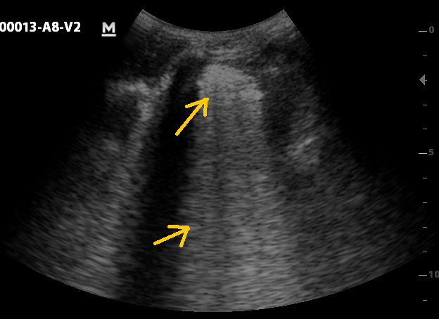
Global Good started by building an algorithm to recognize four key features associated with lung conditions in an ultrasound image – pleural line, B-line, consolidation and pleural effusion. This called for convolutional neural networks (CNNs)—a machine learning method well-suited for image classification tasks. The team trained the algorithm by showing it ultrasound images collected from over 70 pediatric and adult patients. The features were annotated on the images by expert sonographers to ensure accuracy.

Figure 1: Pleural line (upper arrow) and a-lines (lower arrow), indication of normal lung
pneumonia

Figure 2: Consolidation (upper arrow) and merged B-line (lower arrow), indication of abnormal lung fluid and potentially pneumonia

Early tests show that the algorithm can successfully recognize abnormal lung features in ultrasound images and those features can be used to diagnose pneumonia as reliably as X-ray imaging—a highly encouraging outcome.

The algorithm will eventually be installed on an ultrasound device and used by minimally-trained healthcare workers to make high-quality diagnosis accessible to children worldwide at the point of care. Global Good hopes that the device will eventually bring benefits to patients in wealthy markets as well, in the form of a lower-cost, higher quality and faster alternative to X-ray.

4pBA1 – Kidney stone pushing and trapping using focused ultrasound beams of different structure

Oleg Sapozhnikov – olegs@apl.washington.edu
Mike Bailey – bailey@apl.washigton.edu
Adam Maxwell – amax38@uw.edu

Physics Faculty
Moscow State Univerity
Moscow
RUSSIAN FEDERATION

Center for Industrial and Medical Ultrasound
Applied Physics Laboratory
University of Washington
Seattle, WashingtonUNITED STATES

Popular version of paper 4pBA1, “Kidney stone pushing and trapping using focused ultrasound beams of different structure.”
Presented Thursday afternoon, December 1, 2016 at 1:00pmHAST.
172nd ASA Meeting, Honolulu

Urinary stones (such as kidney or bladder stones) are an important health care problem. One in 11 Americans now has urinary stone disease (USD), and the prevalence is increasing. According to a 2012 report from the National Institute of Diabetes and Kidney and Digestive Diseases (Urological Diseases in America), the direct medical cost of USD in the United States is $10 billion annually, making it the most expensive urologic condition.

Our lab is working to develop more effective and more efficient ways to treat stones. Existing treatments such as shock wave lithotripsy or ureteroscopy are minimally invasive, but can leave behind stone fragments that remain in the kidney and potentially regrow into larger stones over time. We have successfully developed and demonstrated the use of ultrasound to noninvasively move stones in the kidney of human subjects. This technology, called ultrasonic propulsion (UP), uses ultrasound to apply a directional force to the stone, propelling it in a direction away from the sonic source, or transducer. Some stones need to be moved towards the ureteropelvic junction (the exit from the kidney that allows stones to pass through the ureter to the bladder) to aid their passage. In other cases, this technology may be useful to relieve an obstruction caused by a stone that may just need a nudge or a rotation to pass, or at least to allow urine to flow and decompress the kidney.

While UP is able to help stones pass, it is limited in how the stones can be manipulated by an ultrasound transducer in contact with the skin from outside the body. Some applications require the stone to be moved sideways or towards the transducer rather than away from it.

To achieve more versatile manipulation of stones, we are developing a new strategy to effectively trap a stone in an ultrasound beam. Acoustic trapping has been explored by several other researchers, particularly for trapping and manipulating cells, bubbles, droplets, and particles much smaller than length of the sound wave. Different configurations have been used to trap particles in standing waves and focused fields. By trapping the stone in an ultrasound beam, we can then move the transducer or electronically steer the beam to move the stone with it.

sapozhnikov1 Kidney stone

Figure 1. The cross section at the focus for the ultrasound pressure of a vortex beam. The pressure magnitude (left) has a donut-shape distribution, whereas the phase (right) has a spiral-shape structure. A stone can be trapped at the center of the ring.

In this work, we accomplished trapping through the use of vortex beams. Typical focused beams create a single region of high ultrasound pressure, producing radiation force away from the transducer. Vortex beams, on the other hand, are focused beams that create a tube-shaped intensity pattern with a spiraling wave front (Fig. 1). The ultrasound pressure in the middle is very low, while the pressure around the center is high. The result is that there is a component of the ultrasound radiation force pushing the stone towards the center and trapping it in the middle of the beam. In addition to trapping, such a beam can apply a torque to the stone and can rotate it.

To test this idea, we simulated the radiation force on spheres of different materials (including stones) to determine how each would respond in a vortex beam. An example is shown in Fig 2. A lateral-axial cross section of the beam is displayed, with a spherical stone off-center in the tube-shaped beam. The red arrow shows that the force on the sphere is away from the center because the stone is outside of the vortex. Once the center of the stone crosses the peak, the force is directed inward. Usually, there is also some force away from the transducer still, but the object can be trapped against a surface.

Figure 2. The simulated tubular field such as occurs in a vortex beam and its force on a stone. In this simulation the transducer is on the left and the ultrasound propagates to the right. The arrow indicates the force which depends on the position of the stone.

We also built transducers and electrically excited them to generate the vortex in experiments. At first, we used the vortex to trap, rotate, and drag an object on the water surface (Fig. 3). By changing the charge of the vortex beam (the rate of spiraling generated by the transducer), we controlled the diameter of the vortex beam, as well as the direction and speed at which the objects rotated. We also tested manipulation of objects placed deep in a water tank. Glass or plastic beads and kidney stones placed on a platform of tissue-mimicking material. By physically shifting the transducer, we were able to move these objects a specified distance and direction along the platform (Fig 4). These results are best seen in videos at apl.uw.edu/pushingstones.

Figure 3. A small object trapped in the center of a vortex beam on the water surface. The ring-shaped impression due to radiation force on the surface can be seen. The phase difference between each sector element of the transducer affects the diameter of the beam and the spin rate. The 2 mm plastic object floating on the surface is made to rotate by the vortex beam.

Figure 4. A focused vortex beam transducer in water (shown on the top) traps one of the styrofoam beads (shown in the bottom) and translates it in lateral direction.

We have since worked on developing vortex beams with a 256-element focused array transducer. Our complex array can electronically move the beam and drag the stone without physically moving the transducer. In a highly focused transducer, such as our array, sound can even be focused beyond the stone to generate an axial high pressure spot to help trap a stone axially or even pull the stone toward the transducer.

There are several ways in which this technology might be useful for kidney stones. In some cases, it might be employed in gathering small particles together and moving them collectively, holding a larger stone in place for fragmentation techniques such as lithotripsy, sweeping a stone when the anatomy inhibits direct radiation force away from the transducer, or, as addressed here dragging or pulling a stone. In the future, we expect to continue developing phased array technology to more controllably manipulate stones. We are also working to develop and validate new beam profiles, and electronic controls to remotely gather the stone and move it to a new location. We expect that this sort of tractor beam could also have applications in manufacturing, such as ever shrinking electronics, and even in space.

This work was supported by RBBR 14-02- 00426, NIH NIDDK DK43881 and DK104854, and NSBRI through NASA NCC 9-58.

References

  1. O.A. Sapozhnikov and M.R. Bailey. Radiation force of an arbitrary acoustic beam on an elastic sphere in a fluid. – J. Acoust. Soc. Am., 2013, v. 133, no. 2, pp. 661-676.
  2. A.V. Nikolaeva, S.A. Tsysar, and O.A. Sapozhnikov. Measuring the radiation force of megahertz ultrasound acting on a solid spherical scatterer. – Acoustical Physics, 2016, v. 62, no. 1, pp. 38-45.
  3. J.D. Harper, B.W. Cunitz, B. Dunmire, F.C. Lee, M.D. Sorensen, R.S. Hsi, J. Thiel, H. Wessells, J.E. Lingeman, and M.R. Bailey. First in human clinical trial of ultrasonic propulsion of kidney stones. – J. Urology, 2016, v. 195, no. 4 (Part 1), pp. 956–964.

3aBA1 – Ultrasound-Mediated Drug Targeting to Tumors: Revision of Paradigms Through Intravital Imaging

Natalya Rapoport – natasha.rapoport@utah.edu
Department of Bioengineering
University of Utah
36 S. Wasatch Dr., Room 3100
Salt Lake City, Utah 84112
USA

Popular version of paper 3aBA1, “Ultrasound-mediated drug targeting to tumors: Revision of paradigms through intravital imaging”
Presented Wednesday morning, May 25, 2016, 8:15 AM in Salon H
171st ASA Meeting, Salt Lake City

More than a century ago, Nobel Prize laureate Paul Ehrlich formulated the idea of a “magic bullet”. This is a virtual drug that hits its target while bypassing healthy tissues. No field of medicine could benefit more from the development of a “magic bullet” than cancer chemotherapy, which is complicated by severe side effects. For decades, the prospects of developing “magic bullets” remained elusive. During the last decade, progress in nanomedicine has enabled tumor-targeted delivery of anticancer drugs via their encapsulation in tiny carriers called nanoparticles. Nanoparticle tumor targeting is based on the “Achilles’ heels” of cancerous tumors – their poorly organized and leaky microvasculature. Due to their size, nanoparticles are not capable to penetrate through a tight healthy tissue vasculature. In contrast, nanoparticles penetrate through a leaky tumor microvasculature thus providing for localized accumulation in tumor tissue.  After tumor accumulation of drug-loaded nanoparticles, a drug should be released from the carrier to allow penetration into a site of action (usually located in a cell cytoplasm or nucleus). A local release of an encapsulated drug may be triggered by tumor-directed ultrasound; application of ultrasound has additional benefits: ultrasound enhances nanoparticle penetration through blood vessel walls (extravasation) as well as drug uptake (internalization) by tumor cells.

For decades, ultrasound has been used only as an imaging modality; the development of microbubbles as ultrasound contrast agents in early 2000s has revolutionized imaging. Recently, microbubbles have attracted attention as drug carriers and enhancers of drug and gene delivery. Microbubbles could have been ideal carriers for the ultrasound-mediated delivery of anticancer drugs.  Unfortunately, their micron-scale size does not allow effective extravasation from the tumor microvasculature into tumor tissue. In Dr. Rapoport’s lab, this problem has been solved by the development of nanoscale microbubble precursors, namely drug-loaded nanodroplets that converted into microbubbles under the action of ultrasound[1-6]. Nanodroplets comprised a liquid core formed by a perfluorocarbon compound and a two-layered drug-containing polymeric shell (Figure 1). An aqueous dispersion of nanodroplets is called nanoemulsion.

Rapoport 1 - Ultrasound-Mediated Drug Targeting

Figure 1. Schematic representation of a drug-loaded nanodroplet

A suggested mechanism of therapeutic action of drug-loaded perfluorocarbon nanoemulsions is discussed below [3, 5, 6]. A nanoscale size of droplets (ca. 250 nm) provides for their extravasation into a tumor tissue while bypassing normal tissues, which is a basis of tumor targeting. Upon nanodroplet tumor accumulation, tumor-directed ultrasound triggers nanodroplet conversion into microbubbles, which in turn triggers release of a nanodroplet-encapsulated drug. This is because in the process of the droplet-to-bubble conversion, particle volume increases about a hundred-fold, with a related decrease of a shell thickness. Microbubbles oscillate in the ultrasound field, resulting in a drug “ripping” off a thin microbubble shell (Figure 2). In addition, oscillating microbubbles enhance internalization of released drug by tumor cells.

Rapoport 2 - Ultrasound-Mediated Drug Targeting

Figure 2. Schematic representation of the mechanism of drug release from perfluorocarbon nanodroplets triggered by ultrasound-induced droplet-to-bubble conversion; PFC – perfluorocarbon

This tumor treatment modality has been tested in mice bearing breast, ovarian, or pancreatic cancerous tumors and has been proved very effective. Dramatic tumor regression and sometimes complete resolution was observed when optimal nanodroplet composition and ultrasound parameters were applied.

3A.Rapoport 3A 3B.Rapoport 3B 3C.Rapoport 3C

(Figure 3. A – Photographs of a mouse bearing a subcutaneously grown breast cancer tumor xenograft treated by four systemic injections of the nanodroplet-encapsulated anticancer drug paclitaxel (PTX) at a dose of 40 mg/kg as PTX. B – Photographs of a mouse bearing two ovarian carcinoma tumors (a) – immediately before and (b) – three weeks after the end of treatment; mouse was treated by four systemic injections of the nanodroplet-encapsulated PTX at a dose of 20 mg/kg as PTX; only the right tumor was sonicated. C – Photographs (a, c) and fluorescence images (b, d) of a mouse bearing fluorescent pancreatic tumor taken before (a, b) and three weeks after the one-time treatment with PTX-loaded nanodroplets at a dose of 40 mg/kg as PTX (c,d). The tumor was completely resolved and never recurred)[3, 4, 6].

In the current presentation, the proposed mechanism of a therapeutic action of drug-loaded, ultrasound-activated perfluorocarbon nanoemulsions has been tested using intravital laser fluorescence microscopy performed in collaboration with Dr. Brian O’Neill (then with Houston Methodist Research Institute, Houston, Texas) [2]. Fluorescently labeled nanocarrier particles (or a fluorescently labeled drug) were systemically injected though the tail vein to anesthetized live mice bearing subcutaneously grown pancreatic tumors. Nanocarrier and drug arrival and extravasation in the region of interest (i.e. normal or tumor tissue) were quantitatively monitored. Various drug nanocarriers in the following size hierarchy were tested: individual polymeric molecules; tiny micelles formed by a self-assembly of these molecules; nanodroplets formed from micelles. The results obtained confirmed the mechanism discussed above.

  • As expected, dramatic differences in the extravasation rates of nanoparticles were observed.
  • The extravsation of individual polymer molecules was extremely fast even in the normal (thigh muscle) tissue; In contrast, the extravasation of nanodroplets into the normal tissue was very slow. (Figure 4. A – Bright field image of the adipose and thigh muscle tissue. B,C – extravasation of individual molecules (B – 0 min; C – 10 min after injection); vasculature lost fluorescence while tissue fluorescence increased. D,E – extravasation of nanodroplets; blood vessel fluorescence was retained for an hour of observation (D – 30 min; E – 60 min after injection).
  • Nanodroplet extravasation into the tumor tissue was substantially faster than that into the normal tissue thus providing for effective nanodroplet tumor targeting.
  • Tumor-directed ultrasound significantly enhanced extravasation and tumor accumulation of both, micelles and nanodroplets (Figure 5). Also, pay attention to a very irregular tumor microvasculature, to be compared with that of a normal tissue shown in Figure 4.
  • The ultrasound effect on nanodroplets was 3-fold stronger than that on micelles thus making nanodroplets a better drug carriers for ultrasound-mediated drug delivery.
  • On a negative side, some premature drug release into the circulation that preceded tumor accumulation was observed. This proposes directions for a further improvement of nanoemulsion formulations.

Rapoport 5 - Ultrasound-Mediated Drug Targeting

Figure 5. Effect of ultrasound on the extravasation of Fluorescence of blood vessels dropped while that of the tumor tissue increased after ultrasound

2aBAa7 – Ultrasonic “Soft Touch” for Breast Cancer Diagnosis

Mahdi Bayat – bayat.mahdi@mayo.edu
Alireza Nabavizadeh- nabavizadehrafsanjani.alireza@mayo.edu
Viksit Kumar- kumar.viksit@mayo.edu
Adriana Gregory- gregory.adriana@mayo.edu
Azra Aliza- alizad.azra@mayo.edu
Mostafa Fatemi- Fatemi.mostafa@mayo.edu
Mayo Clinic College of Medicine
200 First St SW
Rochester, MN 55905

Michael Insana- mfi@illinois.edu
University of Illinois at Urbana-Champaign
Department of Bioengineering
1270 DCL, MC-278
1304 Springfield Avenue
Urbana, IL 61801

Popular version of paper 2aBAa7, “Differentiation of breast lesions based on viscoelasticity response at sub-Hertz frequencies”
Presented Tuesday Morning, May 24, 2016, 9:30 AM, Snowbird/Brighton room
171st ASA Meeting, Salt Lake City

Breast cancer remains the first cause of death among American women under the age of 60. Although modern imaging technologies, such as enhanced mammography (tomosynthesis), MRI and ultrasound, can visualize a suspicious mass in breast, it often remains unclear whether the detected mass is cancerous or non-cancerous until a biopsy is performed.

Despite high sensitivity for detecting lesions, no imaging modality alone has yet been able to determine the type of all abnormalities with high confidence. For this reason most patients with suspicious masses, even those with very small likelihood of a cancer, opt in to undergo a costly and painful biopsy.

It is long believed that cancerous tumors grow in the form of stiff masses that, if found to be superficial enough, can be identified by palpation. The feeling of hardness under palpation is directly related to the tissue’s tendency to deform upon compression.  Elastography, which has emerged as a branch of ultrasound, aims at capturing tissue stiffness by relating the amount of tissue deformation under a compression to its stiffness. While this technique has shown promising results in identifying some types of breast lesions, the diversity of breast cancer types leaves doubt whether stiffness alone is the best discriminator for diagnostic purposes.

Studies have shown that tissues subjected to a sudden external force do not deform instantly, rather they deform gradually over a period of time. Tissue deformation rate reveals another important aspect of its mechanical property known as viscoelasticity. This is the main material feature that, for example, makes a piece of memory foam to feel differently from a block of rubber under the touch. Similar material feature can be used to explore mechanical properties of different types of tissue. In breast masses, studies have shown that biological pathways leading to different breast masses are quite different. While in benign lesions an increase in a protein-based component can potentially increase its viscosity, hence a slower deformation rate compared to normal tissue, the opposite trend occurs in malignant tumors.

In this study, we report on using an ultrasound technique that enables capturing the deformation rate in breast tissue. We studied 43 breast masses in 42 patients and observed that a factor based on the deformation rate was significantly different in benign and malignant lesions (Fig. 1).

The results of this study promise a new imaging biomarker for diagnosis of the breast masses. If such technique proves to be of high accuracy in a large pool of patients, then this technology can be integrated into breast examination procedures to improve the accuracy of diagnosis, reduce unnecessary biopsies, and help detecting cancerous tumors early on

Figure 1 breast cancer

Figure1- Distribution of relative deformation rates for malignant and benign breast lesions. A significantly different relative deformation rates can be observed in the two groups, thus allowing differentiation of such lesions.

2aBAa5 – Sound Waves Helps Assess Bone Condition

Max Denis – denis.max@mayo.edu
507-266-7449

Leighton Wan – wan.leighton@mayo.edu
Matthew Cheong – cheong.matthew@mayo.edu
Mostafa Fatemi – fatemi.mostafa@mayo.edu
Azra Alizad – alizad.azra@mayo.edu
507-254-5970

Mayo Clinic College of Medicine
200 1st St SW
Rochester, MN 55905

Popular version of paper 2aBAa5, “Bone demineralization assessment using acoustic radiation force”
Presented Tuesday morning, May 24, 2016, 9:00 AM in Snowbird/Brighton room
171st ASA Meeting, Salt Lake City, Utah

The assessment of the human skeletal health condition is of great importance ranging from newborn infants to the elderly. Annually, approximately fifty percent of the 550,000 premature newborn infants in the United States suffer from bone metabolism related disorders such as osteopenia, which affect the bone development process into childhood. As we age through adulthood, reductions in our bone mass increases due an unbalance activity in the bone reformation process leading to bone diseases such as osteoporosis; putting a person at risk for fractures in the neck, hip and forearm areas.

Currently bone assessment tools include dual-energy X-ray absorptiometry (DEXA), and quantitative ultrasound (QUS). DEXA is the leading clinical bone quality assessment tool, detecting small changes in bone mineral content and density. However, DEXA uses ionizing radiation for imaging thus exposing patients to very low radiation doses. This can be problematic for frequent clinical visits to monitor the efficacy of prescribed medications and therapies.

QUS has been sought as a nonionizing and noninvasive alternative to DEXA. QUS utilizes measurements of ultrasonic waves between a transmitting and a receiving transducer aligned in parallel along bone surface. Speed of sound (SOS) measurements of the received ultrasonic signal is used to characterize the bone material properties. The determination of the SOS parameter is susceptible to the amount of soft tissue between the skin surface and the bone. Thus, we propose utilizing a high intensity ultrasonic wave known as a “push beam” to exert a force on the bone surface thereby generating vibrations. This will minimize the effects of the soft tissue. The radiate sound wave due to these vibrations are captured and used to analyze the bone mechanical properties.

This work demonstrates the feasibility of evaluating bone mechanical properties from sound waves due to bone vibrations. Under an approved protocol by the Mayo Clinic Institutional Review Board (IRB), human volunteers were recruited to undergo our noninvasive bone assessment technique. Our cohort consisted of clinically confirmed osteopenia and osteoporosis patients, as well as normal volunteers without a history of bone fractures. An ultrasound probe and hydrophone were placed along the volunteers’ tibia bone (Figure 1a). A B-mode ultrasound was used to guide the placement of our push beam focal point onto the bone surface underneath the skin layer (Figure 1b). The SOS was obtained from the measurements.

Denis1 bone

Figure 1. (a) Probe and hydrophone alignment along the tibia bone. (b) Diagram of an image-guided push beam focal point excitation on the bone surface.

In total 14 volunteers were recruited in our ongoing study. A boxplot comparison of SOS between normal and bone diseased (osteopenia and osteoporotic) volunteers in Figure 2, shows that typically sound travels faster in healthy bones than osteoporotic and osteopenia bones with SOS median values (red line) of 3733 m/s and 2566 m/s, respectively. Hence, our technique may be useful as a noninvasive method for monitoring the skeletal health status of the premature and aging population.

Denis2 bone

Figure 2. Normal and bone diseased volunteers sound of speed comparisons.

This ongoing project is being done under an approved protocol by Mayo Institutional Review Board.